|
|
 |
 |
 |
 |
|
|
A 50-year-old man with a long-standing, large-angle exotropia and limitation of adduction in the left eye
Digital Journal of Ophthalmology 2013
Volume 19, Number 4
December 30, 2013
DOI: 10.5693/djo.03.2013.09.004
|
Printer Friendly
Download PDF |
|
|


Reshma A. Mehendale, MD | Department of Ophthalmology, Boston Children’s Hospital, and Harvard Medical School, Boston, Massachusetts Anat O. Stemmer-Rachamimov, MD | Department of Pathology, Massachusetts General Hospital, and Ophthalmic Pathology, Massachusetts Eye and Ear Infirmary, and Harvard Medical School Linda R. Dagi, MD | Department of Ophthalmology, Boston Children’s Hospital, and Harvard Medical School, Boston, Massachusetts
|
|
|
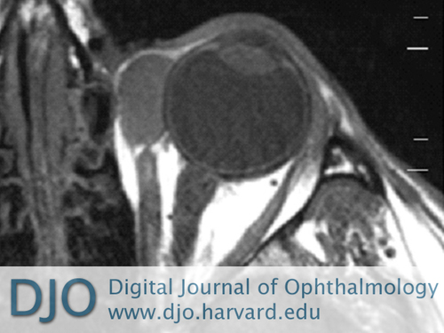
| Ancillary Testing | | Due to the heterogeneous color of the mass, the penetrating vessel, and the lack of adequate history, magnetic resonance imaging (MRI) of the orbits was performed to rule-out malignancy. The MRI showed a well-encapsulated cyst overlying the medial globe, with no evidence of erosion or invasion of surrounding structures. The medial rectus muscle appeared to insert at the posterior pole of the cyst (Figure 2). | |
|
Figure 2
Magnetic resonance imaging of the left orbit showing a well-encapsulated cyst, overlying the medial globe without surrounding erosion; the medial rectus muscle appears to insert at the posterior pole of the cyst.
 |
|
|
 |
 |
 |

|
|
 Welcome, please sign in
  Welcome, please sign in