|
|
 |
|
|
|
|
|
A 56-year-old man with acute vision loss
Digital Journal of Ophthalmology 2016
Volume 22, Number 3
August 18, 2016
DOI: 10.5693/djo.03.2015.05.006
|
Printer Friendly
Download PDF |
|
|
 Fani Akritidou
 | General Hospital of Kavala Maria Karafyloglou | General Hospital of Kavala Dimitrios Karamanis | General Hospital of Kavala
|
|
|
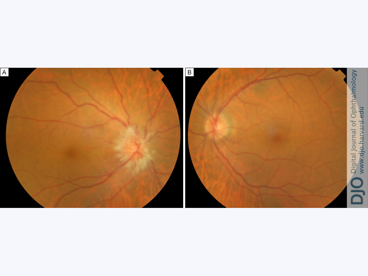
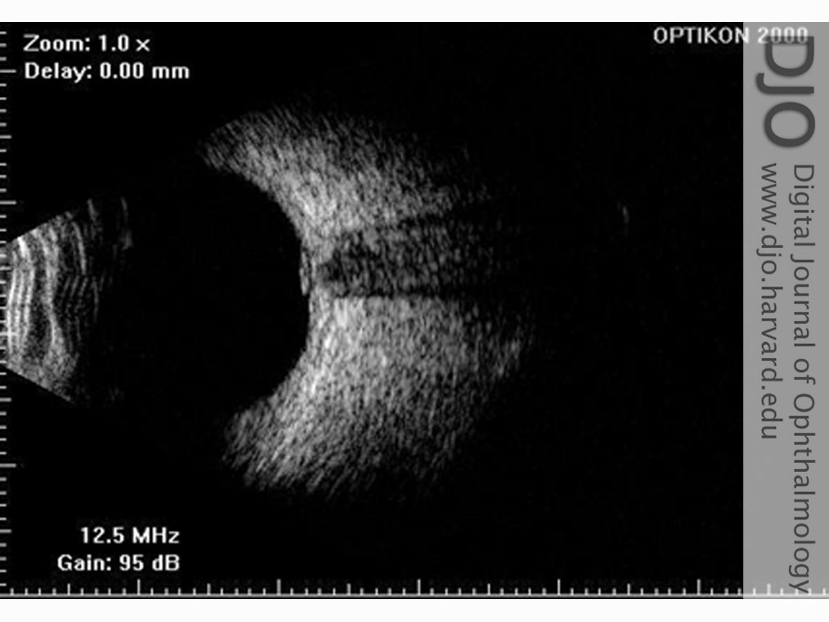
| Examination | On initial examination, the patient’s visual acuity in the right eye was light perception; uncorrected visual acuity in the left eye was 8/10 (Snellen chart at 6 meters; equivalent to 20/25). Intraocular pressure (IOP) measured by Goldmann tonometry was 14 mm Hg in the right eye and 15 mm Hg in the left eye. Ocular motility was normal in both eyes. Anterior segment examination of both eyes was unremarkable. Fundus examination (Figure 1) revealed advanced optic disc edema in the right eye, with no retinal or choroidal lesions; the left eye was normal. Ocular echography (Figure 2) confirmed optic disc swelling in the patient’s right eye.
Neurological examination on admission was normal. Although the patient had no neurological signs or symptoms, he was closely monitored. Two days later, he complained of further visual impairment. On reexamination, visual acuity in his right eye was no light perception, with no other neurological symptomatology. Up to then, all blood analysis and diagnostic tests were normal. On the 4th day after hospitalization, neurological signs and symptoms were apparent, and a lumbar puncture was performed.
On subsequent evaluation, 2 days later, there was further visual loss in the right eye, with no light perception; uncorrected visual acuity in the left eye was stable at 8/10 (20/25). The IOP was 15 mm Hg in both eyes and fundus examination was essentially unchanged. Neurological examination remained normal. On the 4th day of hospitalization the patient complained of severe headache and nausea, and he had epileptic seizures.
| |
|
Figure 1
Fundus photographs of the right eye (A) and left eye (B).
 |
|
|
Figure 2
Ultrasound of the right eye showing the optic disc swelling.
 |
|
|
|
|
|
|
|
 Welcome, please sign in