|
|
 |
|
|
|
|
|
A 66-year-old woman with extensive conjunctival melanosis
Digital Journal of Ophthalmology 2014
Volume 20, Number 2
May 8, 2014
DOI: 10.5693/djo.03.2013.12.002
|
Printer Friendly
|
|
|

Helen Merritt, MD | Orbital Oncology & Ophthalmic Plastic Surgery Program, Department of Plastic Surgery, The University of Texas MD Anderson Cancer Center, Houston, Texas; Ruiz Department of Ophthalmology and Visual Science, The University of Texas Medical School at Houston Mathieu Bakhoum, PhD | Orbital Oncology & Ophthalmic Plastic Surgery Program, Department of Plastic Surgery, The University of Texas MD Anderson Cancer Center, Houston, Texas; University of Texas Medical Branch, Galveston, Texas Matthew Sniegowski, MD | Orbital Oncology & Ophthalmic Plastic Surgery Program, Department of Plastic Surgery, The University of Texas MD Anderson Cancer Center, Houston, Texas Bita Esmaeli, MD | Orbital Oncology & Ophthalmic Plastic Surgery Program, Department of Plastic Surgery, The University of Texas MD Anderson Cancer Center, Houston, Texas
|
|
|
| Examination | On examination, visual acuity was 20/40 in the right eye and 20/30 in the left eye. Intraocular pressure was 6 mm Hg in the right eye and 16 mm Hg in the left eye. The right pupil was noted to be irregular prior to dilation; the left pupil was within normal limits. There was no relative afferent pupillary defect. Extraocular motility was full and visual fields were full to confrontation bilaterally.
Ocular adnexal examination was unremarkable. Slit-lamp examination revealed diffuse pigmentation of the bulbar conjunctiva nearly 360-degrees, with denser pigment superiorly. There was an area of raised conjunctiva superiorly. Pigment did not extend to the palpebral conjunctiva (Figures 1-2). Lids, lashes, cornea, and anterior chambers were unremarkable bilaterally.
Both pupils were pharmacologically dilated. Posterior chamber intraocular lenses were clear and well-positioned in each eye. Fundus examination revealed unremarkable maculae, vessels, and periphery in both eyes. Given the very asymmetric intraocular pressure measurements in the two eyes, the possibility of a trauma-induced, small, self-sealing scleral rupture was considered; nevertheless, given the intact anterior chamber and the extent of abnormal conjunctival pigmentation, a conjunctival biopsy was scheduled.
Two weeks later the patient returned for a scheduled biopsy of the conjunctiva. At this time, examination revealed a corectopic pupil with a peak at 12 o’clock (Figure 3). The conjunctiva was elevated superiorly overlying the area of iris prolapse (Figure 4). Conjunctival pigmentation was present and unchanged from initial examination. | |
|
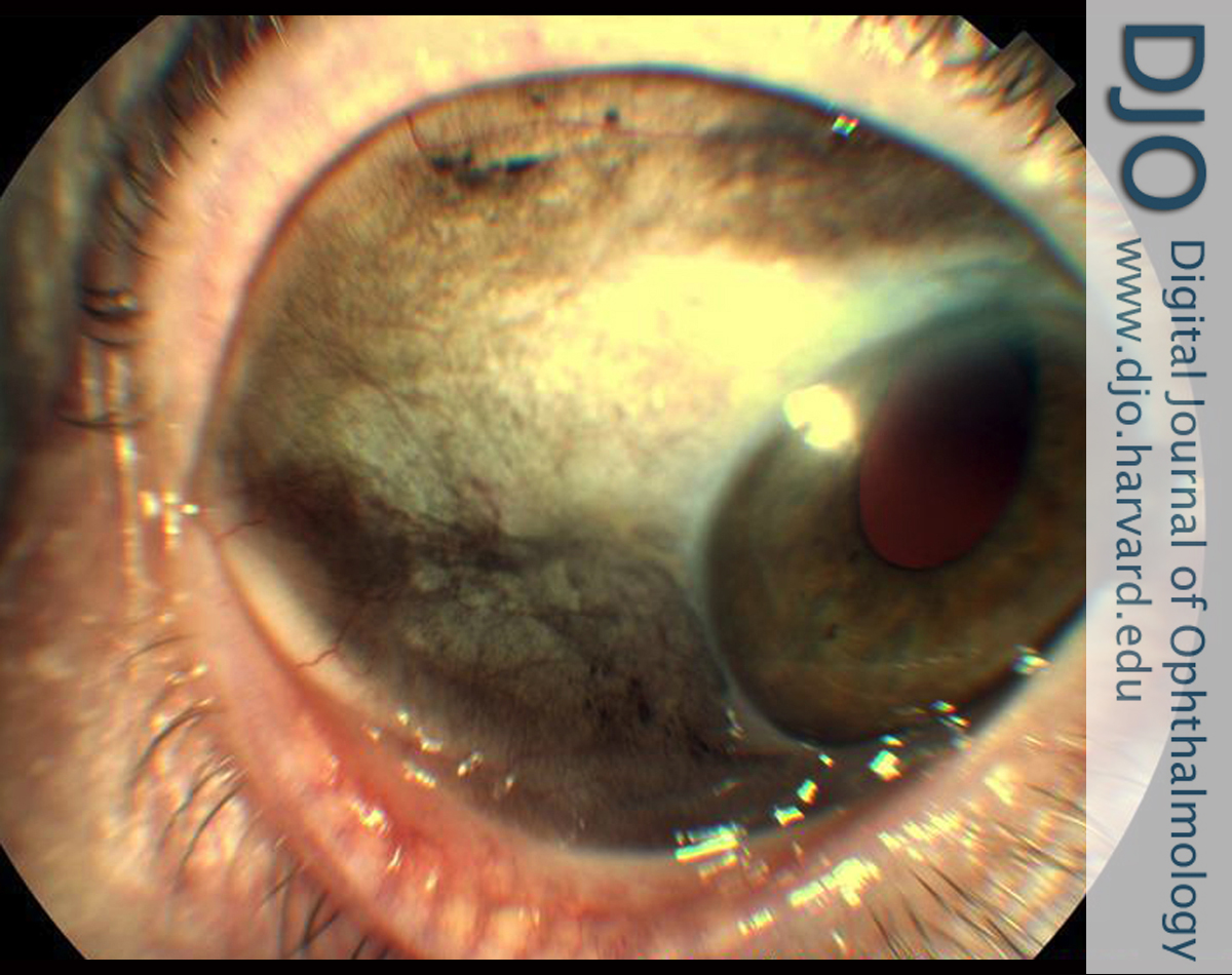
Figure 1
Temporal bulbar conjunctiva with diffuse pigmentation in the patient’s right eye at initial presentation.
 |
|
|
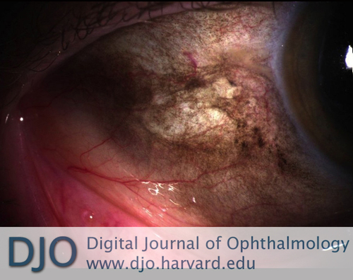
Figure 2
Magnified inferotemporal conjunctiva demonstrating lack of palpebral conjunctival pigment on initial presentation.
 |
|
|
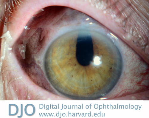
Figure 3
Pupil peak at 12 o’clock on repeat evaluation, 2 weeks following initial presentation.
 |
|
|
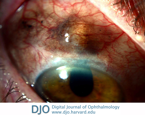
Figure 4
Superior conjunctiva showing iris prolapse with overlying conjunctival pigmentation on repeat evaluation, 2 weeks following initial presentation.
 |
|
|
|
|
|
|
|
 Welcome, please sign in