|
|
 |
|
|
|
|
|
A 48-year-old woman with redness, photophobia, and eye discomfort
Digital Journal of Ophthalmology 2013
Volume 19, Number 2
May 1, 2013
DOI: 10.5693/djo.03.2013.01.003
|
Printer Friendly
Download PDF |
|
|
 Benjamin Erickson, MD
 | Bascom Palmer Eye Institute, Miami, Florida Aleksandra Rachitskaya, MD | Bascom Palmer Eye Institute, Miami, Florida Thomas Shane, MD | Bascom Palmer Eye Institute, Miami, Florida
|
|
|
| Examination | On initial examination, the patient’s uncorrected visual acuity was 20/20 in the right eye and counting fingers in the left eye. Her pupils were 3 mm and reactive with no afferent pupillary defect. Intraocular pressure (IOP) by Goldmann tonometry was 16 mm Hg in the right eye and 31 mm Hg in the left eye. Ocular motility was full in both eyes. The confrontational visual fields were full in the right eye to counting fingers and she was able to detect hand motions in all four quadrants of the left visual field.
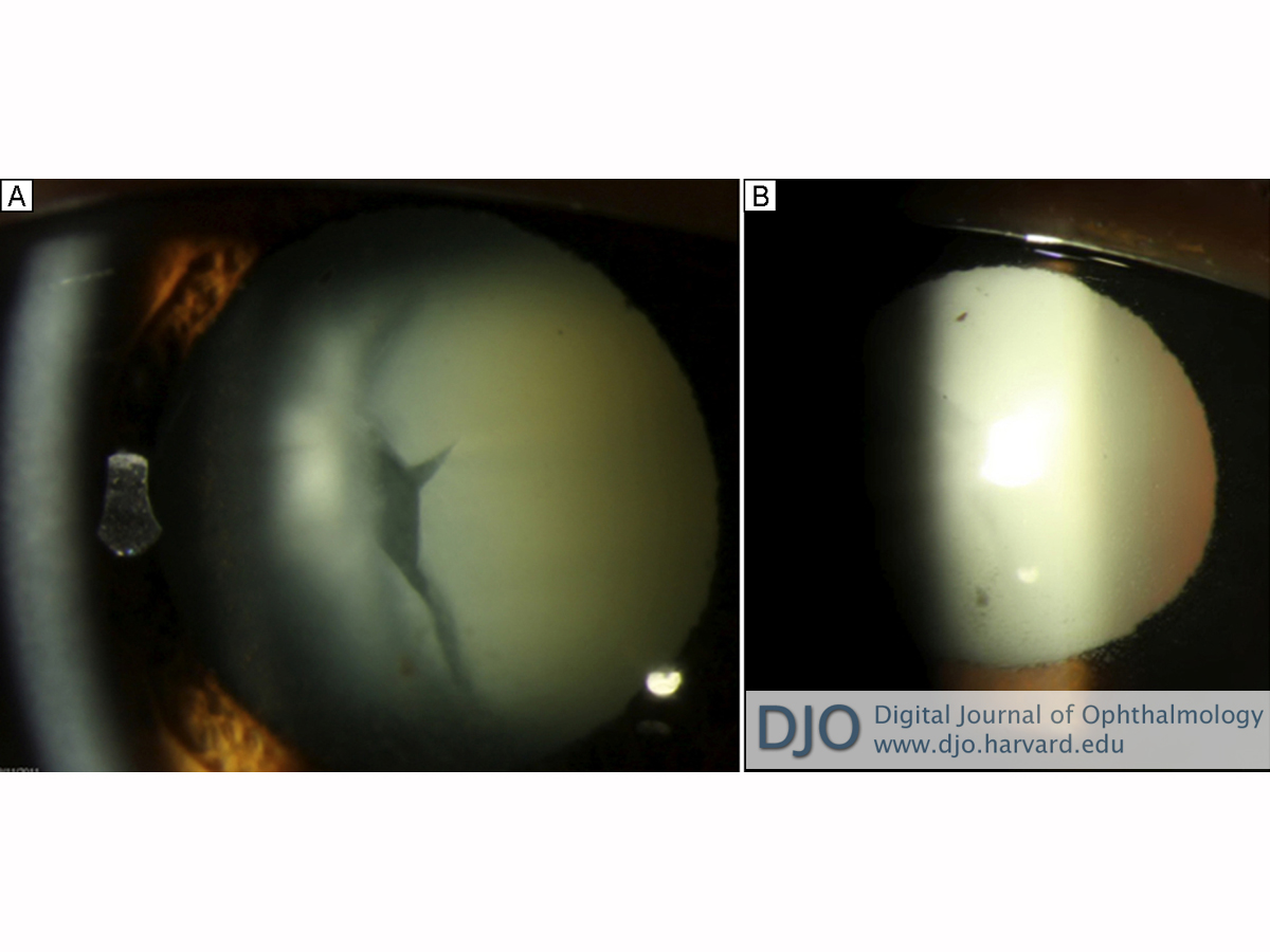
Slit-lamp examination of the left eye was notable for 2+ ciliary injection and for 2+ anterior chamber cell and flare accompanied by scattered mutton fat and stellate keratic precipitates in Arlt’s triangle. No hypopyon was observed. She was noted to have an intumescent cataract with a prominent vertical cleft (Figure 1). There was no anterior bowing of the iris, and the angle was open to the scleral spur without synechiae on gonioscopy.
The anterior segment examination of her right eye was unremarkable. Dilated fundus examination revealed a sharp, pink disc with a 0.4 cup-to-disc ratio and a healthy fundus. There was no posterior view in the left eye due to the density of her cataract. | |
|
Figure 1
A, Intumescent cataract with prominent vertical cleft. B, Retro-illumination of cornea demonstrating stellate and mutton fat keratic precipitates.
 |
|
|
|
|
|
|
|
 Welcome, please sign in