|
|
 |
|
|
|
|
|
A 49-year-old man with unilateral, nontender left eyelid swelling
Digital Journal of Ophthalmology 2014
Volume 20, Number 1
January 17, 2014
DOI: 10.5693/djo.03.2013.09.007
|
Printer Friendly
Download PDF |
|
|
 Brandon J. Wong, BA
 | Keck School of Medicine, University of Southern California, Los Angeles, California Bryan K. Hong, MD | Doheny Eye Institute, Keck School of Medicine, University of Southern California, Los Angeles, California Daman Samrao, MD | Department of Dermatology, Keck School of Medicine, University of Southern California, Los Angeles, California Gene H. Kim, MD | Department of Dermatology, Keck School of Medicine, University of Southern California, Los Angeles, California Narsing A. Rao, MD | Doheny Eye Institute, Keck School of Medicine, University of Southern California, Los Angeles, California
|
|
|
| Diagnosis and Discussion | Given the chronicity of the eyelid edema and lack of response to oral phenylephrine/chlorpheniramine, magnetic resonance imaging (MRI) of the orbits with and without contrast was obtained. The MRI showed globes normal in size, shape, and intensity, with no evidence of intra- or extraconal soft tissue masses and no focal areas of enhancement after contrast. Left periorbital edema consistent with soft-tissue swelling was noted (Figure 3).
The MRI results, combined with a negative laboratory work-up for other possible causes of eyelid edema and questionable tongue abnormality, led to a diagnostic biopsy of the upper left eyelid. A full thickness biopsy, including skin and orbicularis oculi, was taken from the space between the lid crease and the brow, yielding a 3.5 × 1.0 × 0.1 cm tissue specimen. Histopathologic evaluation of the specimen showed diffuse edema of the superficial dermis accompanied by ectatic lymphatics and loosely formed, noncaseating granulomas, many of which appeared to be within the lumens of lymphatics or vessels (Figure 4). All stains, including Gram stain, and special stains for microorganisms (Periodic acid-Schiff and acid-fast bacilli) were negative, and no polarizable foreign material was identified. No eosinophils were noted. Immunohistochemical staining with D2-40 and CD68 confirmed the presence of ectatic lymphatics and intralymphatic histiocytosis. Based on the clinical symptoms, combined with the histopathologic results, a diagnosis of Melkersson-Rosenthal syndrome was made.
Melkersson-Rosenthal syndrome (MRS) is classically described as a triad of recurrent facial paralysis, orofacial edema, and fissuring of the tongue (lingua plicata).(2) The dominant feature of the syndrome is orofacial edema, which is painless, nonpitting, unilateral, and typically includes the eyelids and lips.(3) The facial paralysis can be unilateral (mimicking Bell’s palsy) or bilateral.(4) Lingua plicata is the rarest component of the triad and is not considered pathognomonic of the disease. Patients may be mono- or oligo-symptomatic, and in such cases the presence of only one or two features in the triad can make this a challenging diagnosis. Moreover, the features may present over a period of years, which further complicates the diagnosis.(5) The etiology of the syndrome remains unclear, though autoimmune or viral causes have been hypothesized.(3,6) Symptoms tend to present around the second or third decade of life, although infrequently they can present in childhood or after 50 years of age.(7) Studies regarding the gender predisposition for the syndrome are conflicting, with females being equally or slightly more affected than males.(3,8)
Our patient did not manifest the classic triad of Melkersson-Rosenthal syndrome, presenting only with isolated left upper eyelid edema and no clinical evidence of facial nerve paralysis or gross lingua plicata. A diagnosis of MRS requires a high index of clinical suspicion as well as histopathological evidence for confirmation of the diagnosis. The distinctive histopathology of MRS is described as non-caseating, granulomatous lymphangitis with granulomas within and around the lymphatics and blood.(3,4,8) MRS should be considered in patients presenting with eyelid edema of unknown etiology and biopsy should be performed.(9)
Initial treatment for Melkersson-Rosenthal syndrome usually consists of topical, intralesional, or systemic corticosteroid therapy, although the syndrome is often refractory to treatment. Patients with MRS are also treated with nonsteroidal, anti-inflammatory drugs such as metronidazole, dapsone, acyclovir, methotrexate, and thalidomide, with varying levels of response.(5) Clofazimine, an anti-lepromatous drug, has also been reported to decrease some of the eyelid swelling and associated granulomas.(10)
Surgical management of eyelid edema, such as debulking procedures or blepharoplasty, may be indicated if the edema is unresponsive to steroids, if it becomes visually significant, or becomes cosmetically unacceptable for the patient.(11) | |
|
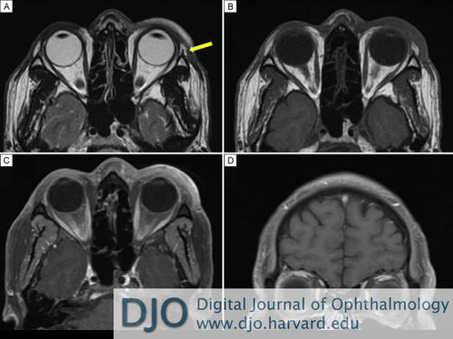
Figure 3
Magnetic resonance imaging of the orbits obtained in sequence. A, Axial T2-weighted imaging before contrast showing left eyelid tissue swelling and no evidence of lacrimal gland enlargement (yellow arrow). B, Axial T1-weighted imaging before contrast showing left eyelid soft tissue swelling. C, Axial T1-weighted imaging after contrast with fat suppression showing continued left eyelid soft tissue swelling and no focal areas of enhancement. D, Coronal T1-weighted imaging after contrast with fat suppression showing no extraorbital masses or abnormal enlargement of extraocular muscles.
 |
|
|
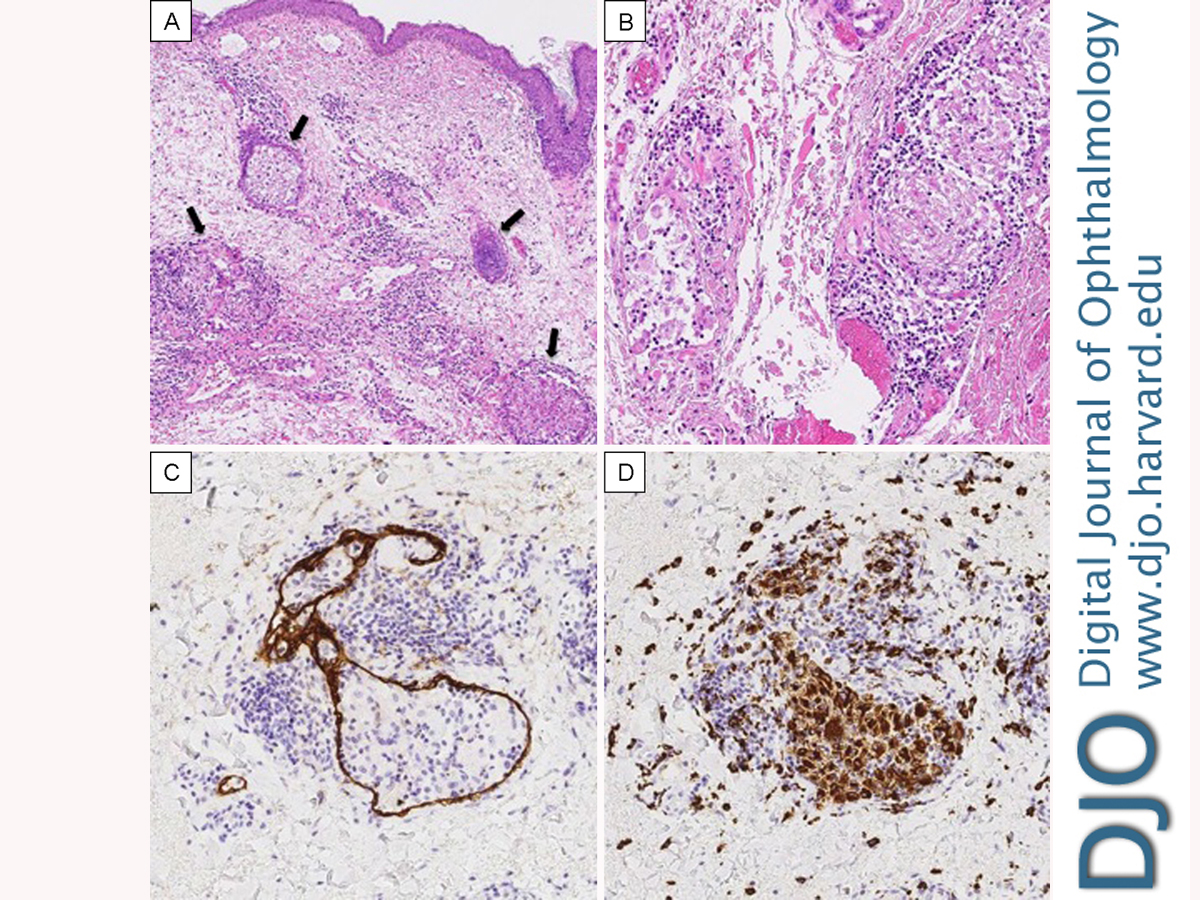
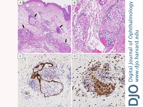
Figure 4
A, A low-power photomicrograph showing dermal edema, dilated lymphatics and noncaseating granulomas (black arrows) associated with the lymphovasculature along with an infiltrate of chronic inflammatory cells (hematoxylin and eosin, original magnification × 10). B, High-power image of the granulomas showing a loose collection of histiocytes possibly within the lumen of a lymphatic or vessel. Chronic inflammation is present surrounding the granulomas (hematoxylin and eosin, original magnification × 20). C, The D2-40 stain highlights the presence of a lymphatic wall surrounding a loosely formed granuloma. D, The presence of the intralymphatic granuloma is confirmed by the CD68 stain.
 |
|
|
|
|
|
|
|
 Welcome, please sign in