|
|
 |
|
|
|
|
|
A 5-year-old girl with left upper eyelid swelling
Digital Journal of Ophthalmology 2012
Volume 18, Number 4
December 31, 2012
DOI: 10.5693/djo.03.2012.05.001
|
Printer Friendly
Download PDF |
|
|
 Syed Shoeb Ahmad, MBBS, MS, FAEH, FCLI
 | Department of Ophthalmology, Queen Elizabeth Hospital, Kota Kinabalu, Malaysia Shuaibah Abdul Ghani, MS | Department of Ophthalmology, Queen Elizabeth Hospital, Kota Kinabalu, Malaysia Khoo Say Peng, MS | Dr Peter Kong Specialist Eye Centre, Kota Kinabalu, Malaysia Puliventhan Sellamuthu, MS | Department of Neurosurgery, Queen Elizabeth Hospital, Kota Kinabalu, Malaysia
|
|
|
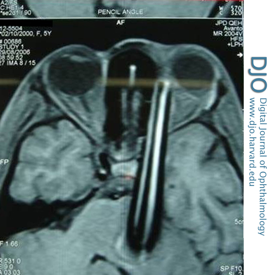
| Ancillary Testing | | Computed tomography (CT) revealed an intraorbital foreign body. The hospital neurosurgeon was consulted and a magnetic resonance imaging (MRI) scan was requested. The MRI revealed a long, linear orbital foreign body extending from the left orbit through the medial part of the temporal lobe up to the brain stem (Figure 2). When confronted with the MRI report, the patient’s mother continued to deny any injury with a sharp object. Based on the general condition of the patient, absence of neurological deficits and radiological findings, the neurosurgeon did not recommend an angiography. | |
|
Figure 2
Magnetic resonance imaging showing the intraorbital foreign body extending to the brainstem.
 |
|
|
|
|
|
|
|
 Welcome, please sign in