|
|
 |
|
|
|
|
|
A 64-year-old woman with dilated right pupil, nausea, and headache
Digital Journal of Ophthalmology 2013
Volume 19, Number 1
January 27, 2013
DOI: 10.5693/djo.03.2012.11.001
|
Printer Friendly
Download PDF |
|
|
 Ali Haider, BMedSc, MBBS
 | The Canberra Hospital, Canberra, Australia Prashanth J. Rao, MBBS, MS | The Canberra Hospital, Canberra, Australia
|
|
|
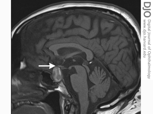
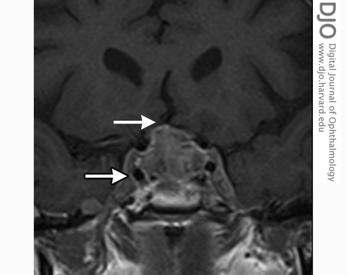
| Ancillary Testing | | Computed tomography (CT) of the head demonstrated an intrasellar mass abutting the optic chiasm, with no evidence of subarachnoid hemorrhage or an unruptured aneurysm on CT angiogram. Magnetic resonance imaging (MRI) of the brain confirmed a pituitary macroadenoma with suprasellar extension and hemorrhage with mass effect and tumor extension into the pituitary infundibulum and cavernous sinuses (Figures 2-3). Endocrine studies showed no evidence of anterior pituitary dysfunction except reduced level of thyroid-stimulating hormone (TSH), suggestive of a response to long-term thyroxine. Urinary cortisol was elevated (272; normal <150 nmol/day). | |
|
Figure 2
T1-weighted magnetic resonance imaging (MRI), sagittal view, showing hyperintense sellar mass (optic chiasm indicated by arrow).
 |
|
|
Figure 3
MRI, coronal view, showing gross expansion of pituitary fossa with heterogeneous high T1 signal reflecting blood products. There is infiltration of the cavernous sinuses (outlined arrow) bilaterally with effacement of the undersurface of the optic chiasm (arrow).
 |
|
|
|
|
|
|
|
 Welcome, please sign in


