|
|
 |
|
|
|
|
|
A 44-year-old woman with a 3-month history of bilateral, painless visual loss in the absence of other symptoms
Digital Journal of Ophthalmology 2012
Volume 18, Number 4
December 31, 2012
DOI: 10.5693/djo.03.2012.12.001
|
Printer Friendly
Download PDF |
|
|
 Emily Shao, MBBS, BSC
 | Department of Ophthalmology, Chelsea and Westminster Hospital, London, UK Kevin Gallagher, BMBCh | Department of Ophthalmology, Chelsea and Westminster Hospital, London, UK Nabeel Malik, MBBS, FRCOPHTH | Department of Ophthalmology, Chelsea and Westminster Hospital, London, UK
|
|
|
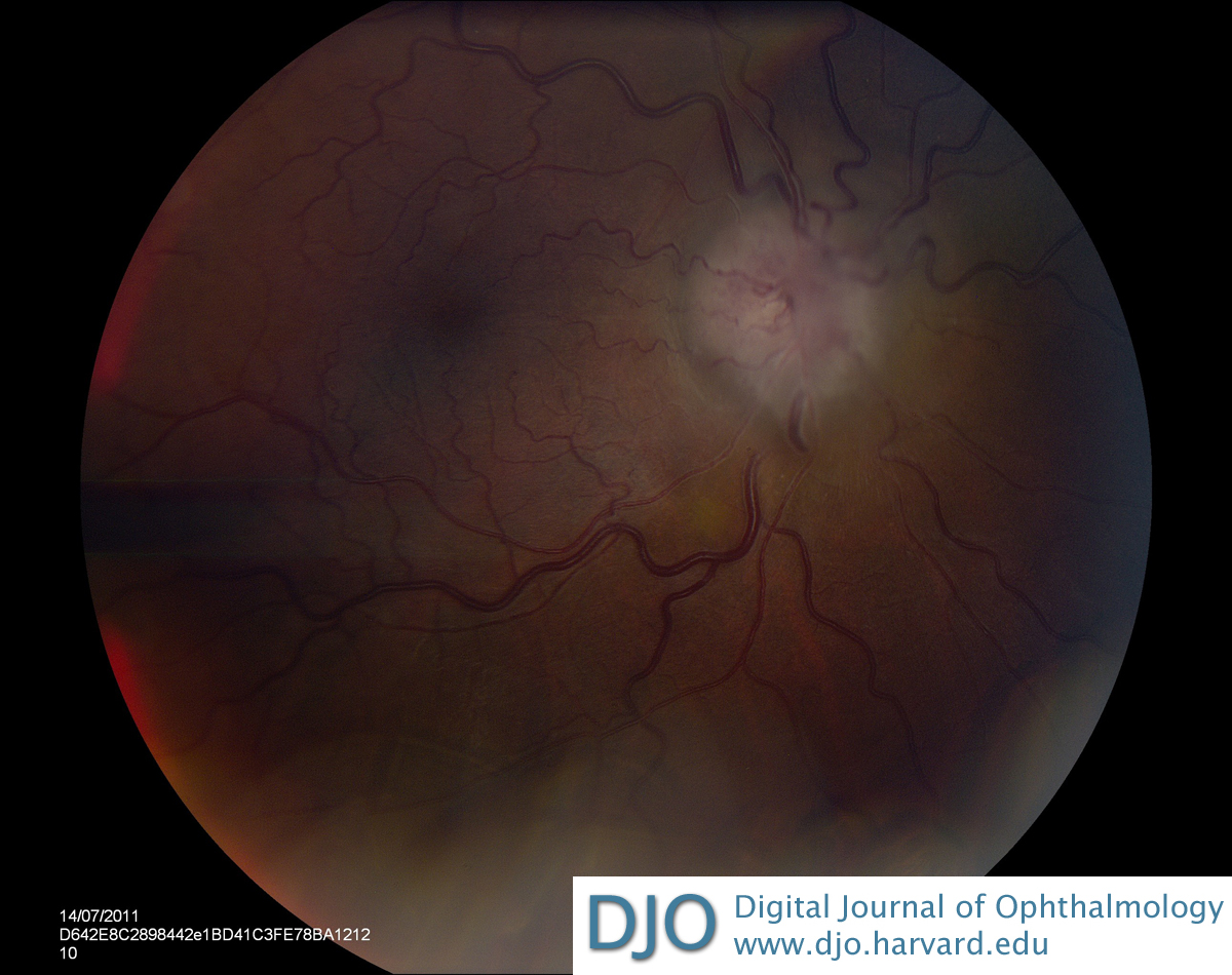
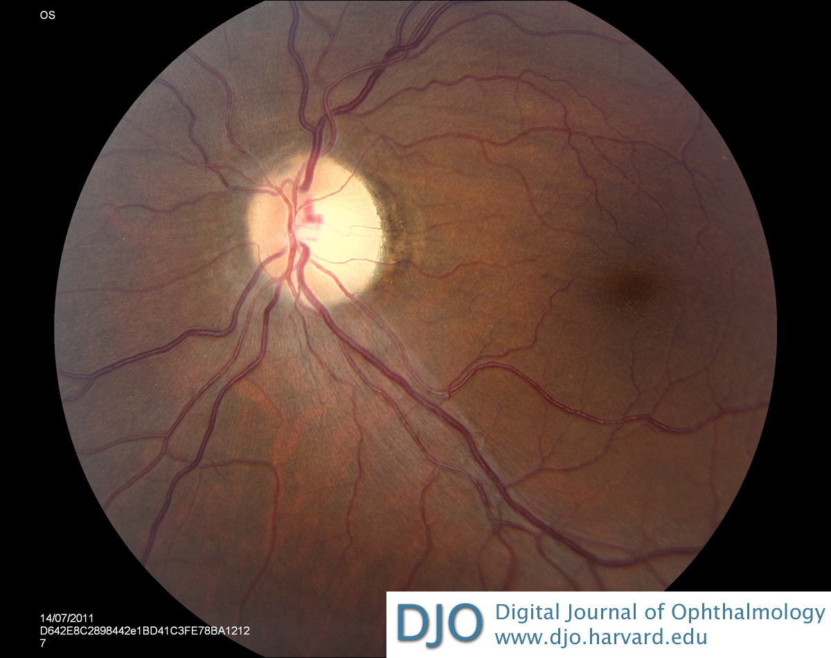
| Examination | | On examination, visual acuity was hand movements in the right eye and no light perception (NLP) in the left eye. There was no relative afferent papillary defect. Pupils were round and reactive to light. The anterior segments were normal. On dilated fundus examination, she was found to have a grossly swollen right optic disc (Figure 1) and a pale left optic disc (Figure 2). | |
|
Figure 1
Papilledema of the right optic disc.
 |
|
|
Figure 2
Left optic disc pallor.
 |
|
|
|
|
|
|
|
 Welcome, please sign in