|
|
 |
|
|
|
|
|
A 44-year-old woman with a 3-month history of bilateral, painless visual loss in the absence of other symptoms
Digital Journal of Ophthalmology 2012
Volume 18, Number 4
December 31, 2012
DOI: 10.5693/djo.03.2012.12.001
|
Printer Friendly
Download PDF |
|
|
 Emily Shao, MBBS, BSC
 | Department of Ophthalmology, Chelsea and Westminster Hospital, London, UK Kevin Gallagher, BMBCh | Department of Ophthalmology, Chelsea and Westminster Hospital, London, UK Nabeel Malik, MBBS, FRCOPHTH | Department of Ophthalmology, Chelsea and Westminster Hospital, London, UK
|
|
|
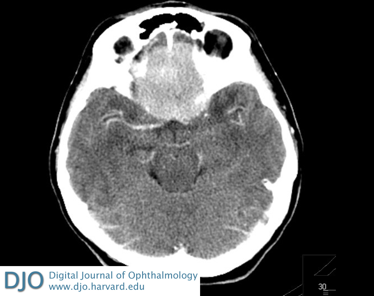
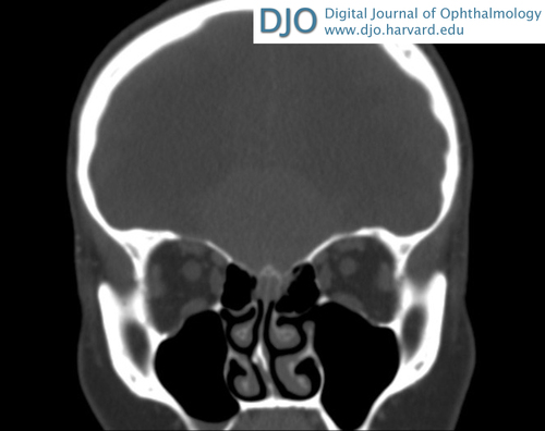
| Ancillary Testing | | Computed tomography (CT) of the head and orbits revealed a frontal mass extending into the planum sphenoidale and the pituitary fossa with the appearance of a large anterior cranial fossa floor meningioma (Figure 3). No abnormality was seen in the globes, optic nerves, extraocular muscles, or any intraconal structure in either orbit (Figure 4). | |
|
Figure 3
Computed tomography (CT) of the head showing a well-circumscribed homogenous lesion (4.6 × 4.4 × 2.5 cm) in the midline of the frontal region with surrounding edema consistent with meningioma.
 |
|
|
Figure 4
CT of the orbits showing no abnormality involving the globes, optic nerves, extraocular muscles, or any intraconal structures in either orbit; an intracranial mass is seen in the frontal region consistent with meningioma in the coronal view.
 |
|
|
|
|
|
|
|
 Welcome, please sign in