|
|
 |
|
|
|
|
|
A 39-year-old woman with unilateral metamorphosias
Digital Journal of Ophthalmology 2011
Volume 17, Number 4
November 20, 2011
DOI: 10.5693/djo.03.2011.11.001
|
Printer Friendly
Download PDF |
|
|
 Lígia Ribeiro
 | Department of Ophthalmology, Centro Hospitalar Vila Nova de Gaia, Portugal Sidnei Barge | Department of Ophthalmology, Centro Hospitalar Vila Nova de Gaia, Portugal Luís Silva | Department of Ophthalmology, Centro Hospitalar Vila Nova de Gaia, Portugal Arnaldo Brandão | Department of Ophthalmology, Centro Hospitalar Vila Nova de Gaia, Portugal Dália Meira | Department of Ophthalmology, Centro Hospitalar Vila Nova de Gaia, Portugal
|
|
|
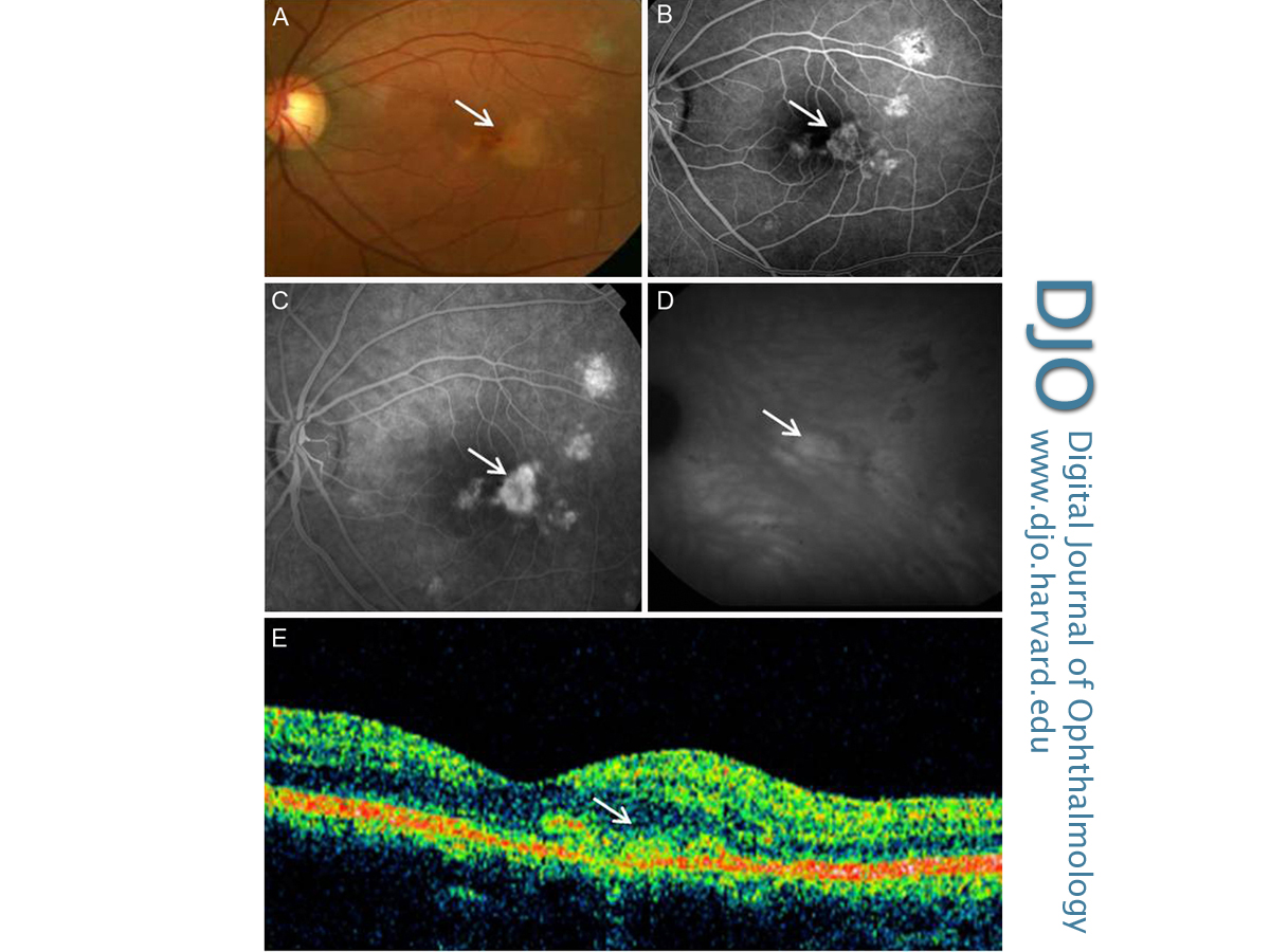
| Ancillary Testing | Fluorescein angiography showed well-demarcated, small, areas of early- and late-phase hyperfluorescence (Figure 1B). The larger macular lesion was hyperfluorescent, with blurred contours and markedly increased hyperfluorescence over the course of the imaging consistent with a juxtafoveal choroidal neovascular membrane (Figure 1C).
Indocyanine green angiography showed several areas of hypofluorescence in the right eye (Figure 1D), which corresponded to the visible subretinal lesions observed during fundus examination; the juxtafoveal lesion was hyperfluorescent, which confirmed the activity of the neovascular membrane.
Time-domain optical coherence tomography revealed a macular thickness of 246 µm in the left eye; there was no subretinal fluid (Figure 1E).
A complete work-up, including chest X-ray, serology for syphilis, immunoglobulins (IgM and IgG) for Toxoplasma and Borrelia burgdorferi and angiotensin-converting enzyme levels, was normal. | |
|
Figure 1
Left eye of patient with juxtafoveal choroidal neovascularization (arrows) secondary to punctate inner choroidopathy. A, Fundus photograph. B, Fluorescein angiogram showing early phase hyperfluorescence. C, Fluorescein angiogram showing midvenous phase hyperfluorescence. D, Indocyanine green angiogram showing areas of hyperfluorescence. E, Optical coherence tomography (OCT).
 |
|
|
|
|
|
|
|
 Welcome, please sign in