|
|
 |
|
|
|
|
|
A 16-year-old girl with bilateral optic disc swelling
Digital Journal of Ophthalmology 2011
Volume 17, Number 1
March 12, 2011
DOI: 10.5693/djo.03.2011.02.003
|
Printer Friendly
Download PDF |
|
|
|
|
|
|
| Ancillary Testing | To exclude all causes of hypertension in the patient’s age group, the pediatricians performed a detailed work-up, including urea and electrolytes, urinary cortisol measurements (9 AM and midnight), liver function tests, gamma glutamyl transferase, urine HCG, full blood count, erythrocyte sedimentation rate, C-reactive protein, thyroid function tests, and early morning urine test for microalbuminuria. All tests were normal.
A lumbar puncture showed clear and colorless cerebrospinal fluid, total protein count of 23 mg/100 mL (normal, 15-60 mg/100 mL), gamma globulin 11% of the total protein (normal, 3%-12% of total protein), glucose level of 76 mg/100 mL (normal, 50-80 mg/100mL), cell count of 0-5 white blood (all mononuclear) cells (normal, 0-5) with no red blood cells, and chloride level of 117 mEq/L (normal, 116-130 mEq/L). Microbiology of the cerebrospinal fluid was unremarkable, with no growth.
An electrocardiogram was normal.
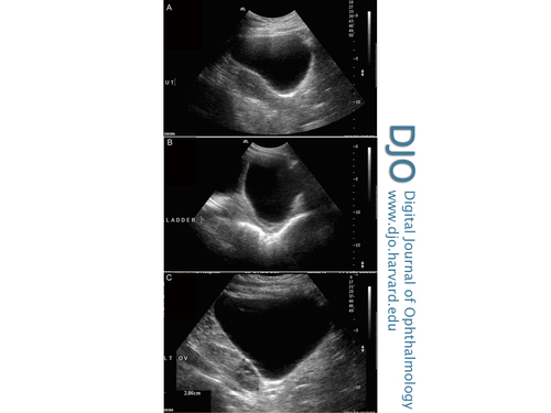
In view of the normal lab work-up, an ultrasound of the abdomen was requested to rule out gynecological or renal causes of hypertension. Ultrasonography of the abdomen showed a large abdominal cyst extending from the symphysis pubis to the level of xyphisternum (Figure 3). Magnetic resonance imaging of the abdomen showed a cyst arising from the pelvis and measuring 34 × 29 × 12 cm. Within its right side border an additional septated area was seen, suggesting a small additional follicular cyst within a large ovarian cyst. No renal compression or hydronephrosis was noted. | |
|
Figure 3
Ultrasonography of the abdomen showing a large abdominal cyst extending from symphysis pubis to the level of xyphisternum, approximately 25 cm in trasnsverse, 40-60 cm in longest axis and 20 cm deep (all at deepest and widest points): in relation to the uterus (UT) (A), the bladder (LADDER) (B), and the left ovary (LT OV) (C). Content is generally clear and anechoic, with a suggestion of a trace of debris at its dependant aspect.
 |
|
|
|
|
|
|
|
 Welcome, please sign in


