|
|
 |
|
|
|
|
|
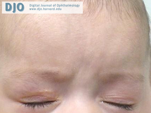
A 7-week-old infant with right upper eyelid mass
Digital Journal of Ophthalmology 2010
Volume 16, Number 4
October 2, 2010
DOI: 10.5693/djo.03.2010.09.001
|
Printer Friendly
Download PDF |
|
|

John Davis | University of Texas Medical Branch at Galveston Kapil Kapoor | University of Texas Medical Branch at Galveston
|
|
|
| Treatment | The patient’s family was presented with all treatment options and made aware of the concern for occlusional amblyopia given the rapid progression of the mass. Propranolol was not advised by the primary ICU team due to concerns regarding a heart murmur and the patient’s overall cardiac status. After a full discussion of the indications, risks, benefits, and alternatives, surgical excision of the right upper eyelid mass was performed (Figure 2A-D). Pathology slides are shown in Figure 3A-D.
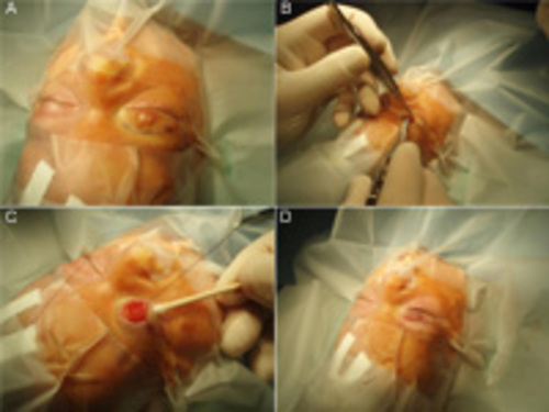
Twelve days postoperatively, the patient was noted to have a recurrence, which continued to progress over the next two weeks. At that point (1 month postoperatively), the patient was given an intralesional corticosteroid injection (50/50 mix of 40 mg/mL triamcinolone and 6 mg/mL betamethasone). There was no recurrence at 2 months’ follow-up (Figure 4).
| |
|
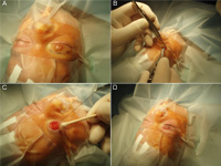
Figure 2
Surgical excision of right upper eyelid lesion. A, Preoperative patient preparation, with incision marked above eyelid crease. B, Incision made along previously noted mark. C, Exposure of eyelid mass. D, Postoperative photograph after mass excision and closure.
 |
|
|
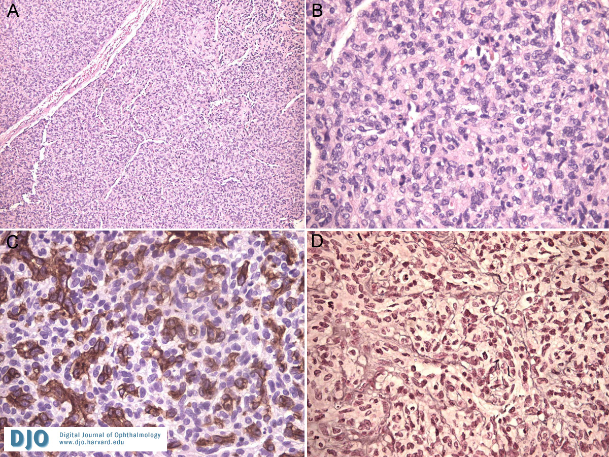
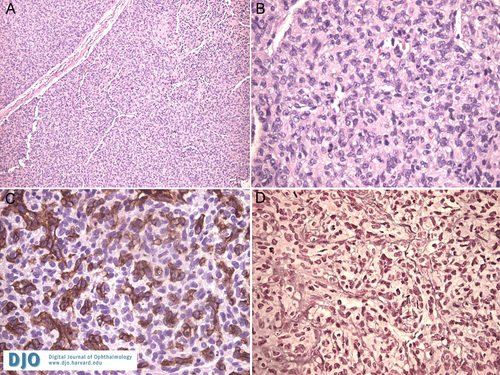
Figure 3
A, Histology demonstrating high cellularity with only rare vascular components (hematoxylin and eosin, original magnification ×40). B, On higher magnification, numerous mitotic figures were noted, reflecting a highly proliferative lesion (hematoxylin and eosin, ×200). C, A CD31 stain emphasizing the numerous endothelial channels in the lesion (×200). D, Positive reticulin stain surrounding capillary vessels (×200).
 |
|
|
Figure 4
Patient 2 months postoperatively. Note the surgical scar visible on the right upper eyelid above the lid crease. There is no evidence of recurrence.
 |
|
|
|
|
|
|
|
 Welcome, please sign in