|
|
A 37-year-old man with a black spot in his vision
Digital Journal of Ophthalmology 2010
Volume 16, Number 1
February 9, 2010
DOI: 10.5693/djo.03.2009.12.001
|
Printer Friendly
Download PDF |
|
|
|
|
|
|
| Examination |
The visual acuity was 20/20 in both eyes. The intraocular pressure was 14 mm Hg in both eyes. The pupillary examination was normal, and there was no dyschromatopsia in either eye. Anterior segment exam was normal. Visual fields showed mild global constriction in both eyes. Dilated fundus exam (Figure 1) and fluorescein angiogram (FA) (Figure 2) showed mild vasculitis in both eyes and an area of arterial occlusion in the right eye.
The patient had a repeat examination 2 weeks later following worsening of neurologic symptoms. Examination at this time showed visual acuity of 20/20 in both eyes. The pupil exam was normal. There was significant superior visual field constriction in both eyes on confrontation. Dilated fundus exam showed large areas of retinal nonperfusion, multiple areas of arterial occlusion, and active vasculitis in both eyes (Figure 3). Fluorescein angiogram confirmed multiple areas of active vasculitis in both eyes (Figure 4).
|
|
|
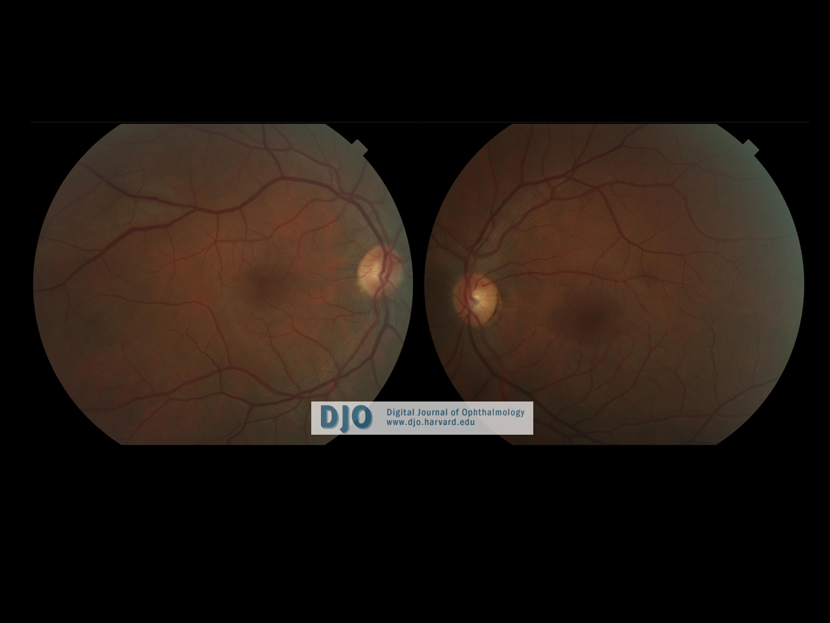
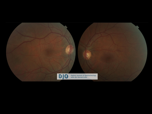
Figure 1
Fundus photos showing posterior pole OU on initial ophthalmologic exam.
 |
|
|
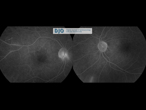
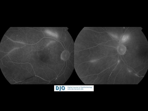
Figure 2
Fluorescein angiography demonstrating vascular occlusion OD and mild vasculitis OU.
 |
|
|
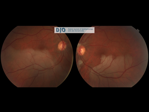
Figure 3
Fundus photos on repeat examination 2 weeks after initial exam showing significant areas of retinal vascular occlusions in both eyes with large areas of retinal nonperfusion OU and cotton wool spots OS.
 |
|
|
Figure 4
Fluorescein angiography demonstrating significant arteriolar occlusions OU and active retinal vasculitis OU.
 |
|
 Welcome, please sign in