|
|
 |
|
|
|
|
|
A 48-year-old man presents with bilateral corneal deposits
Digital Journal of Ophthalmology 2009
Volume 15, Number 4
November 27, 2009
|
Printer Friendly
Download PDF |
|
|
|
|
|
|
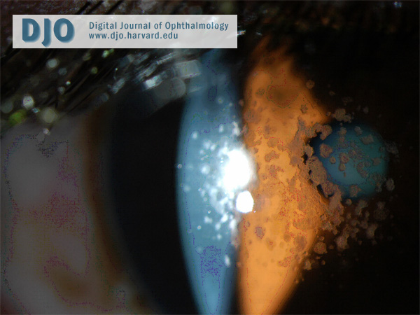
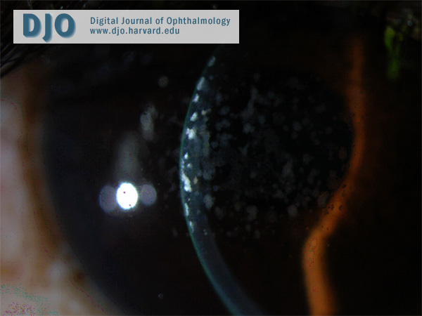
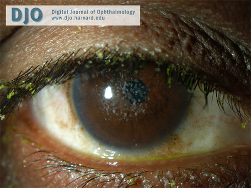
| Examination | | On examination, his best-corrected visual acuity was 20/20 in his right eye and 20/30 in his left eye. The pupils were equally reactive with no afferent pupillary defect. Slit-lamp examination (see Figures 1-3) was significant for bilateral small, discrete, sharply demarcated, grayish-white opacities in the anterior central stroma with intervening clear areas. Notably, there was no conjunctival inflammation. The sclera was white and quiet. The anterior chamber was deep and quiet in both eyes. Tonometry and Schirmer’s test were normal in both eyes. On funduscopic examination, the optic disc, macula and vessels were within normal limits in both eyes. | |
|
Figure 1
External photo of the right eye.
 |
|
|
Figure 2
In type 2 granular corneal dystrophy, the “bread crumb” lesions coalesce into fewer larger ring-shaped granular deposits with sharply defined intervening clear zones.
 |
|
|
Figure 3
Slit-lamp photograph demonstrating anterior stromal location of the deposits.
 |
|
|
|
|
|
|
|
 Welcome, please sign in