|
|
 |
|
|
|
|
|
A 68-year-old man with a recurrent orbital lesion
Digital Journal of Ophthalmology 2009
Volume 15, Number 4
September 2, 2009
|
Printer Friendly
Download PDF |
|
|
 Marc-Andre Rheaume, MD
 | University of Montreal Guy Allaire, MD, FRCPC | University of Montreal Akram Rahal, MD, FRCSC | University of Montreal Vijayabalan Balasingam, MD, FRCSC, PhD | University of Montreal Patrick R. Boulos, MD, FRCSC | University of Montreal
|
|
|
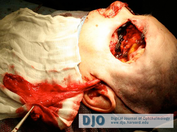
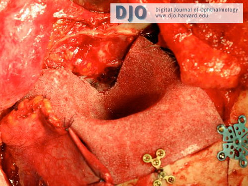
| Treatment | A radical exenteration of the left orbit including the roof, lateral wall and orbital apex was performed as a combined procedure by otolaryngology, neurosurgery and oculofacial surgery (Figure 4). All margins were free of residual tumor on intraoperative frozen sections. The left orbit was reconstructed using a Medpor™ Complete Orbital implant and a 2-part temporalis muscle and temporoparietal fascial flap with skin grafts. The implant was contoured to fit the defect and was fixed to the surrounding bone using plate and screws (Figure 5).
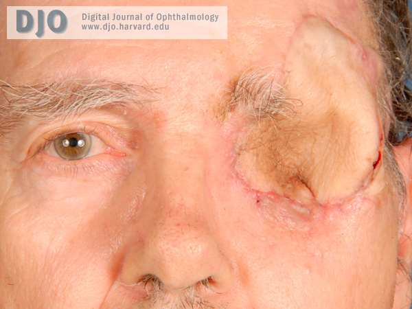
One week later the patient developed necrosis of the distal portion of his temporoparietal flap and of the overlying skin graft. This exposed the Medpor™ implant. The patient was taken back to the operating room and the area was debrided and reconstructed with a radial forearm free flap. A left superficial parotidectomy with facial nerve dissection and a left cervical neck dissection were also performed since a tumor was found in the ipsilateral parotid gland on reviewing the MRI. All were free of malignancy. Surprisingly, an entirely benign pleomorphic adenoma was found in the ipsilateral parotid gland with no evidence of metastasis. The patient then received radiotherapy to the orbital region. Figure 6 shows him 3 months after the second procedure. | |
|
Figure 4
Perioperative image showing the extent of the defect.
 |
|
|
Figure 5
Orbit reconstructed with the MedporTM implant.
 |
|
|
Figure 6
Picture taken 3 months after the last surgery.
 |
|
|
|
|
|
|
|
 Welcome, please sign in