Welcome, please sign in
Follow DJO on Facebook Follow DJO on Twitter
Grand Rounds
  Most Recent Cases
  Dates of Case
  Type of Case
  Submit a Grand Round.
  Register with DJO to receive personalized updates.

If you're already a
member, please sign in.
A 68-year-old man with a recurrent orbital lesion
Digital Journal of Ophthalmology 2009
Volume 15, Number 4
September 2, 2009
Printer Friendly

Download PDF


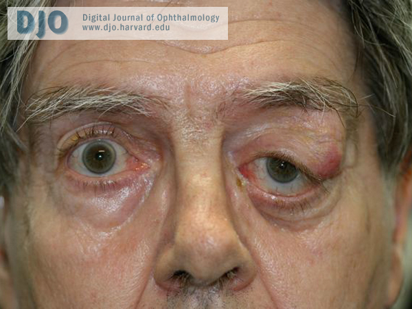
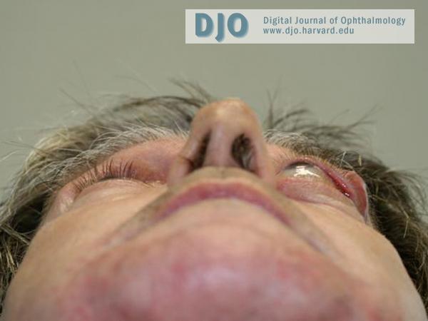
Marc-Andre Rheaume, MD | University of Montreal
Guy Allaire, MD, FRCPC | University of Montreal
Akram Rahal, MD, FRCSC | University of Montreal
Vijayabalan Balasingam, MD, FRCSC, PhD | University of Montreal
Patrick R. Boulos, MD, FRCSC | University of Montreal
Examination
On initial examination, best corrected visual acuity was 20/40 in the right eye and 20/30 in the left eye. Pinhole testing improved visual acuity to 20/25 in both eyes. Color vision tested with the HRR plates was normal bilaterally. A painful and fixed mass in the left superotemporal orbit was identified on palpation and skin changes were seen overlying it. It was also found to displace the globe inferonasally (Figure 1a). Hertel exophtalmolometry measurements were 15 mm in the right eye and 21 mm in the left eye (Figure 1b). Extraocular movements were full on the right side with a slight limitation of abduction in the left eye. Pupils were normally reactive with no afferent papillary defect. Slit-lamp examination of the anterior and posterior segments was entirely normal. No choroidal folds were seen in the left eye. Evaluation of the fifth cranial nerve revealed a diminished sensitivity in the left ophthalmic division. The maxillary division was normal bilaterally. No abnormal cervical lymph nodes were palpated.
top
Figure 1a
External examination revealed a fixed mass that was painful on palpation as well as left hypoglobus.

Figure 1b
Photograph demonstrating proptosis of the left globe.