|
|
 |
|
|
|
|
|
A 68-year-old man with a recurrent orbital lesion
Digital Journal of Ophthalmology 2009
Volume 15, Number 4
September 2, 2009
|
Printer Friendly
Download PDF |
|
|
 Marc-Andre Rheaume, MD
 | University of Montreal Guy Allaire, MD, FRCPC | University of Montreal Akram Rahal, MD, FRCSC | University of Montreal Vijayabalan Balasingam, MD, FRCSC, PhD | University of Montreal Patrick R. Boulos, MD, FRCSC | University of Montreal
|
|
|
| Ancillary Testing | Radiographic Studies
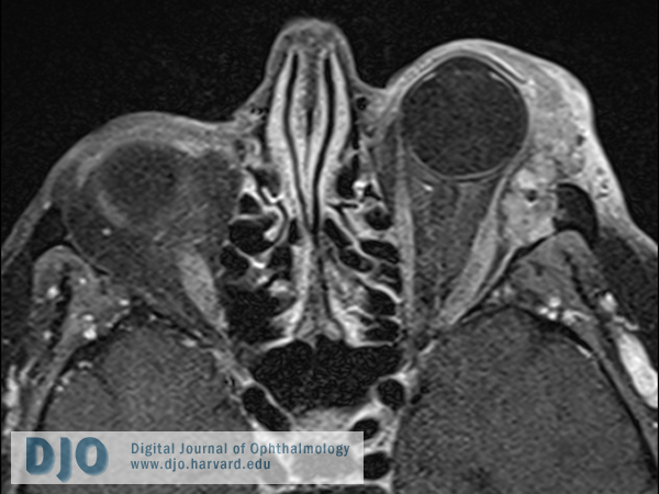
An orbital MRI was obtained. A polylobulated mass in the superolateral quadrant of the left orbit was found. There was apparent destruction of the lateral wall and lateral part of the orbital roof by the tumor. Upper eyelid infiltration was also seen. The globe itself was normal. (Figure 2a-b)
Pathology
An incisional biopsy was performed via an anterior orbitotomy. It revealed the presence of a malignant mixed tumor arising from a pleomorphic adenoma. Immunohistochemical studies with keratin 7 were also obtained and confirmed the presence of epithelial cells in the malignant clone. (Figure 3a-b)
A consultation with an oncologist was requested and an extensive systemic workup including cerebral MRI, PET scan, chest x-ray, abdominopelvic CT scan, CBC, renal and liver tests was ordered. All results were within normal limit. The remainder of the complete physical examination was normal. | |
|
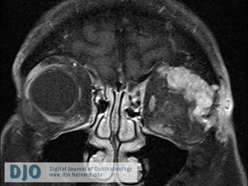
Figure 2a
T1-weighted fat suppressed image showing the ill-defined mass in the left orbit. The lacrimal gland cannot be distinguished.
 |
|
|
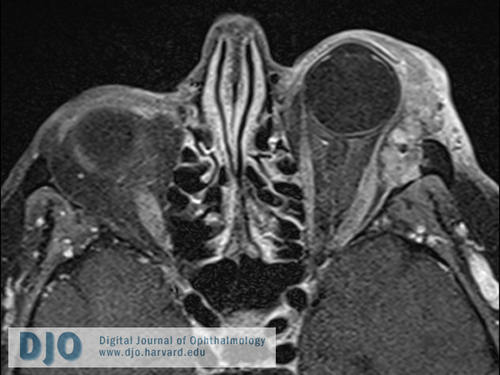
Figure 2b
T1-weighted fat suppressed image showing the tumor coming in close contact with the left lateral rectus muscle and its insertion on the globe. The optic nerve is intact. The lesion measures 5.1 x 3.4 x 2.4 cm.
 |
|
|
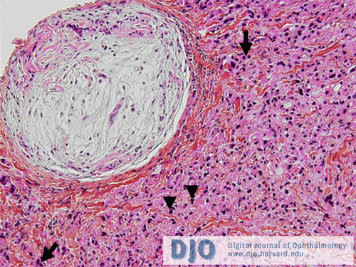
Figure 3a
Benign mixed tumor composed of clumps of ovoid and fusiform-shaped cells embedded in a myxoid stroma is seen in the upper left corner. The malignant component is on the right. Mitotic figures can be seen (arrows) as well as dense chromatin in the nuclei of cells showing an elevated nuclear-cytoplasmic ratio (arrowheads).
 |
|
|
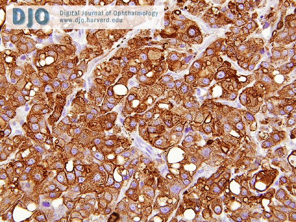
Figure 3b
Positive staining of the malignant cells for keratin 7, which confirms the epithelial origin of the tumor.
 |
|
|
|
|
|
|
|
 Welcome, please sign in