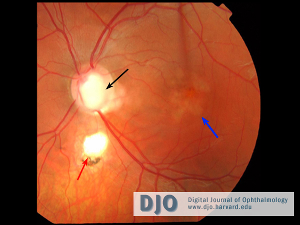
The patient was emmetropic with acuities of 6/6 (20/20) in the right eye and 6/9 (20/30) in the left eye. He was N5 (Jaeger 1) in both eyes and had full color vision on Ishihara plate testing. The anterior segments were normal in both eyes. On fundoscopic examination, the right eye was normal, and the left eye had a large diameter optic disc with a coloboma, an inferior retinochoroidal coloboma, central retinal pigment epithelial atrophy and macular retinal elevation (Figure 1).
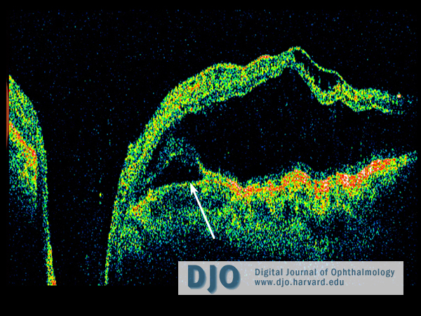
Ocular coherence tomography (OCT) of the left eye revealed schisis-like separation of the retinal layers in the macula with a partial outer layer detachment adjacent to the disc and strands of tissue bridging the schisis cavity (Figure 2). A connection between the inner layer separation and the optic disc was suggested. Mean central 1 mm retinal thickness was 697 microns. Visual acuity deteriorated 2 months after presentation to 6/18 (20/60).
Figure 1
Color fundus photograph of the left eye showing the colobomatous disc (black arrow), retino-choroidal coloboma (red arrow), foveal retinal pigment epithelial atrophy and macular elevation (blue arrow).
Figure 2
Linear horizontal OCT image pre-treatment through the disc. The scan shows schisis-like separation of the retinal layers in the macula with a partial outer layer detachment adjacent to the disc (arrow).
 Welcome, please sign in