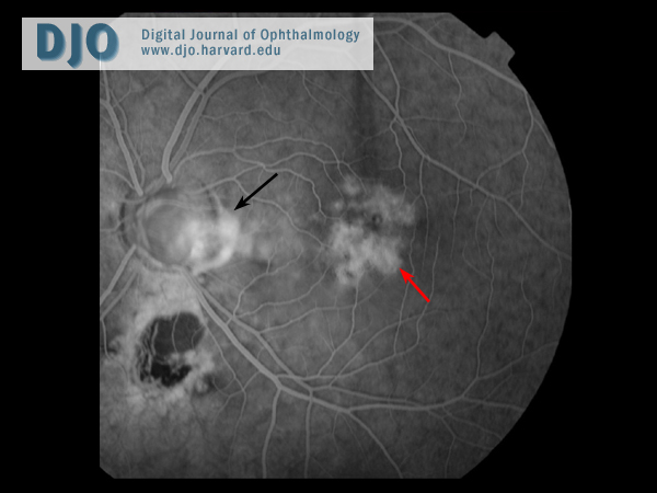
Fluorescein angiography of the left eye revealed late hyperfluorescence within the disc coloboma and window defects between the optic disc and the fovea (Figure 3).
Figure 3
Fluorescein angiogram 33 seconds. There is leakage within the coloboma and between the disc and the fovea (black arrow). Patchy retinal pigment epithelial atrophy can be seen around the fovea (red arrow).
 Welcome, please sign in