|
|
 |
|
|
|
|
|
A 25-month-old girl with vision loss, nystagmus, and anomalous head posture
Digital Journal of Ophthalmology 2009
Volume 15, Number 1
February 23, 2009
|
Printer Friendly
Download PDF |
|
|
 Joel Metzger, MD
 | Senior Flight Surgeon, Training Air Wing Six, Pensacola, FL, USA Richard Hertle, MD | Children's Hospital of Pittsburgh, Pittsburgh, PA, UPMC Eye Center, Eye & Ear Institute, Pittsburgh, PA, and University of Pittsburgh School of Medicine, Pittsburgh, PA, USA John Avallone, MD | Ophthalmology Associates of Greater Annapolis Edward Cheeseman, M.D. | University of South Carolina School of Medicine, Charleston, SC and Uniformed Services University of the Health Sciences, Bethesda, MD, USA
|
|
|
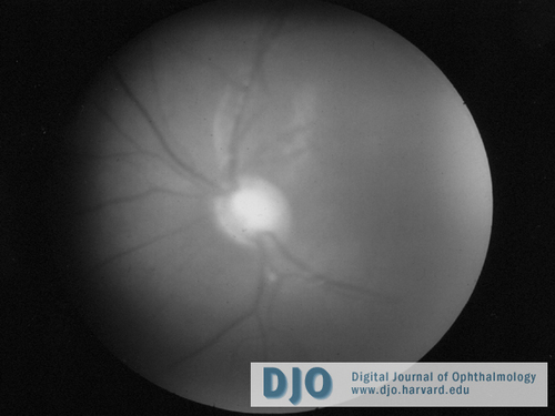
| Examination | Initial exam revealed an alert, cooperative, age-appropriate 25-month-old girl with a left head turn of approximately 30-40 degrees. The patient was noted to have central, unsteady, and unmaintained fixation OU. The pupils were of equal size without an afferent pupillary defect. During an MRI under sedation, intraocular pressures (IOP) were 32-36 mmHg OD and 30-38 mmHg OS. Her corneal diameters were 12.5 mm OU without evidence of edema, infiltrate, or neovascularization. Haab’s striae ran longitudinally in the inferior corneas of both eyes. On gonioscopy, the angle appeared anomalous with a high anterior insertion of a flat iris. The lenses and vitreous were clear OU. Dilated fundoscopic exam demonstrated a 0.9 X 0.9 cup-to-disc ratio OD and 0.9 X 0.8 cup-to-disc ratio OS. Both nerve heads showed pallor and deep cupping. An image of the left nerve is shown in Figure 1. Her refraction was -9.00 +2.00 x180 OD and –7.00 +2.50 x180 OS. The MRI was normal. Combination timolol-dorzolamide ophthalmic drops were initiated, trabeculotomy was scheduled, and the patient was referred for further evaluation of her nystagmus.
Additional examination revealed full versions and ductions and a comitant esotropia. The patient had a left AHP, preferring right gaze. She had variable, symmetric, conjugate, small-to-moderate amplitude, and moderate-to-high frequency multiplanar involuntary ocular oscillations with a null position in right gaze of 30-40 degrees. There was no latent component and saccades, pursuit, and vestibulo-ocular reflexes were otherwise normal. | |
|
Figure 1
Fundus photograph of left optic nerve showing extreme cupping and atrophy present in both nerves at the time of diagnosis.
 |
|
|
|
|
|
|
|
 Welcome, please sign in


