|
|
 |
|
|
|
|
|
A 34-year-old man with visual complaints and a tapetal-like reflex
Digital Journal of Ophthalmology 2008
Volume 14, Number 14
July 28, 2008
|
Printer Friendly
|
|
|
 Joao C.M.L. Ribeiro
 | Massachusetts Eye and Ear Infirmary Cynthia S. Chiu | Massachusetts Eye and Ear Infirmary Christine Ament | Massachusetts Eye and Ear Infirmary John I. Loewenstein | Massachusetts Eye and Ear Infirmary
|
|
|
| Examination | External examination demonstrated hypertelorism and shallow orbits. Best corrected visual acuity was counting fingers in the right eye and 20/40 in the left eye. The intraocular pressure was 16 mm Hg in the right eye and 15 mm Hg in the left. The pupils were round, equal and reactive to light. A right afferent pupillary defect was detected. Color testing could not be done in the right eye due to poor acuity. The patient had normal Farnsworth D15 results in the left eye. The patient was only able to read the control on Ishihara plates. Slit-lamp examination was unremarkable.
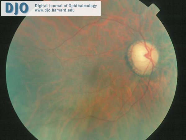
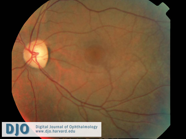
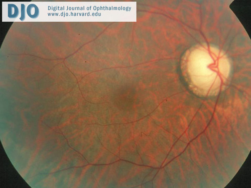
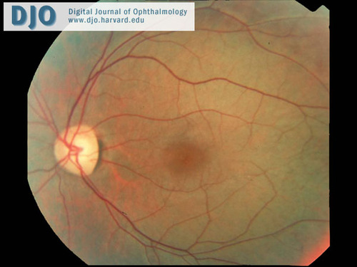
Dilated fundus examination showed pallor and significant cupping of the optic disc in the right eye. There was a striking yellow-gold sheen to the posterior retina (Figure 1). In the left eye, the disc was normal. There was a similar, but more marked, yellow-gold sheen that extended anterior to the vascular arcades (Figure 3). The sheen was less marked in both eyes after 1 hour of dark adaptation (Figures 2 and 4). | |
|
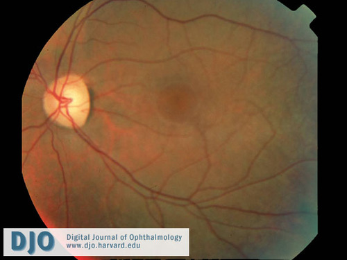
Figure 1
Fundus photo of the right eye. The dilated examination showed pallor and significant cupping of the optic disc. While there were no definite abnormalities observed in the macula, a striking yellow-gold sheen to the retina was observed, particularly posteriorly.
 |
|
|
Figure 2
The tapetal-like sheen in the right eye was less marked after one hour of dark adaptation.
 |
|
|
Figure 3
Fundus photo of the left eye. A normal disc and unremarkable macula are noted, but there is a marked golden-yellow sheen to the fundus extending beyond the arcades.
 |
|
|
Figure 4
The tapetal-like sheen of the left fundus was less marked after one hour of dark adaptation.
 |
|
|
|
|
|
|
|
 Welcome, please sign in