Joao C.M.L. Ribeiro | Massachusetts Eye and Ear Infirmary Cynthia S. Chiu | Massachusetts Eye and Ear Infirmary Christine Ament | Massachusetts Eye and Ear Infirmary John I. Loewenstein | Massachusetts Eye and Ear Infirmary
Electroretinography (ERG)
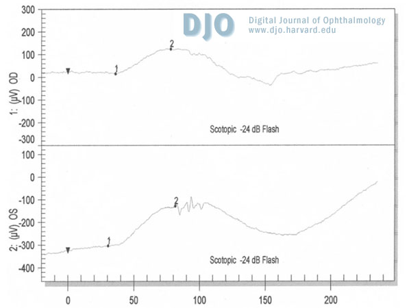
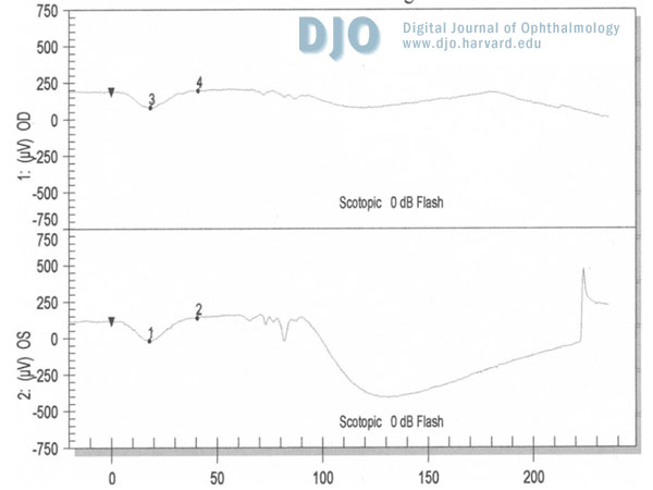
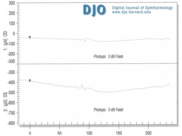
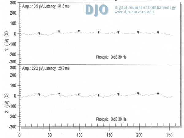
A full-field ERG was performed according to the international standard protocol. Results were compared to a database of healthy individuals or patients with no abnormal ERG values obtained in our lab. The patient’s ERG showed a reduced dim scotopic response in the right eye and a normal result in the left eye (Figure 5). The bright scotopic b-wave amplitude was reduced in both eyes but implicit times were normal (Figure 6). The bright photopic single flash response was non-recordable in both eyes (Figure 7). The 30Hz flicker responses were severely reduced in both eyes (Figure 8). The implicit time was borderline in the right eye and normal in the left eye.
Humphrey Visual Field (HVF) testing:
The left visual field showed severe constriction. The test could not be performed in the right eye.
 Welcome, please sign in