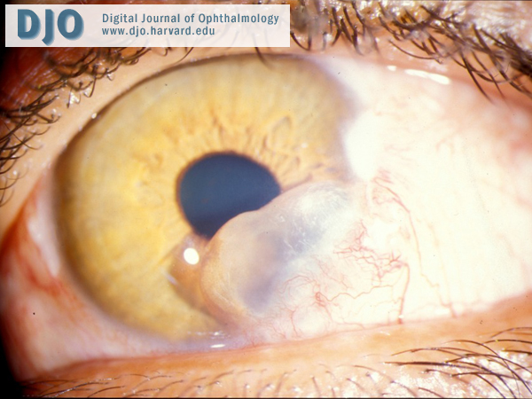
Visual acuity was 20/30 OS, and IOP was 14 mm Hg OS. Slit-lamp examination revealed a 5.0 mm x 3.5 mm cystic lesion from 3:00 to 6:00 in the left eye (Figure 1). There was an underlying transillumination defect of the iris. There was no cell or flare present in the anterior chamber. The lens was clear, and dilated fundus exam was unremarkable with no clinical evidence of macular edema.
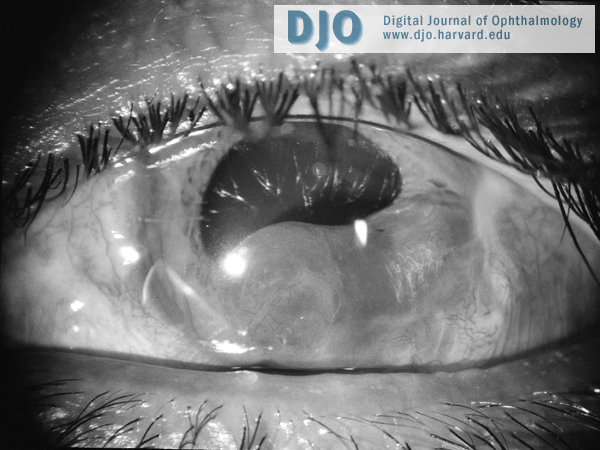
The patient was treated with cycloplegia and asked to return in 1 month, at which time the visual acuity had fallen to 20/70. IOP was still in the mid-teens. The cyst had enlarged to 8.0 mm x 4.5 mm and was encroaching on the visual axis (Figure 2). The decline in vision was attributed to this encroachment on the visual axis, as there was no evidence of any other anterior segment or fundus abnormalities. Cycloplegia was continued to provide the patient with a larger pupil. He was asked to return in 1 month for ultrasound biomicroscopy and re-examination.
At the next visit, visual acuity was stable at 20/70, and IOP was 17 mm Hg. The cyst now measured 10.0 mm x 8.0 mm and appeared to be obscuring the visual axis (Figure 3).
 Welcome, please sign in