Welcome, please sign in
Follow DJO on Facebook Follow DJO on Twitter
Grand Rounds
  Most Recent Cases
  Dates of Case
  Type of Case
  Submit a Grand Round.
  Register with DJO to receive personalized updates.

If you're already a
member, please sign in.
A 23-year-old man with a cystic iris lesion
Digital Journal of Ophthalmology 2008
Volume 14, Number 15
August 1, 2008
Printer Friendly


Glenn W. Thompson | William Beaumont Hospital
Steven P. Dunn | Michigan Cornea Consultants
Ancillary Testing
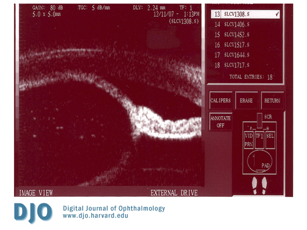
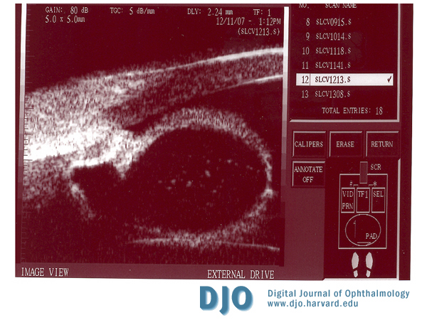
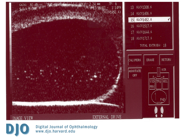
Ultrasound biomicroscopy (UBM) revealed an iris cyst originating from the original wound. The cyst appeared to be adherent to the surrounding cornea. Figure 4 shows the cyst separating the anterior and posterior layers of the iris. Figure 5 shows the extent of the cyst near the ciliary body; note this image is from the peripheral portion of the cyst, since at the wound the differentiation of cyst and ciliary body was difficult to view. Figure 6 shows the full extent of the cyst in the anterior-posterior direction, which measured 3.80 microns.
top
Figure 4
UBM showing cyst walls made up of anterior and posterior iris.

Figure 5
UBM centered near ciliary body.

Figure 6
UBM showing anterior-posterior extent of cyst.