|
|
 |
|
|
|
|
|
A 24-year-old woman with blurred vision and eye pain
Digital Journal of Ophthalmology 2008
Volume 14, Number 16
August 18, 2008
|
Printer Friendly
|
|
|
 Aristeidis Konstantinidis
 | Royal United Hospital Bath, UK Ioannis Athanasiadis | Milton Keynes General Hospital, UK Nikolaos Kozeis | Hippokarateio General Hospital , Greece Claire Workmann | Coventry & Warwickshire University Hospital, UK Yajati Ghosh | Coventry & Warwickshire University Hospital, UK
|
|
|
| Ancillary Testing | An intravenous fluorescein angiogram was normal.
Blood work revealed a normal B12 level, negative antinuclear antibody (ANA) level, negative anti-double stranded DNA antibody level, and normal angiotensin converting enzyme level. Chest x-ray showed no evidence of interstial lung disease or hilar adenopathy.
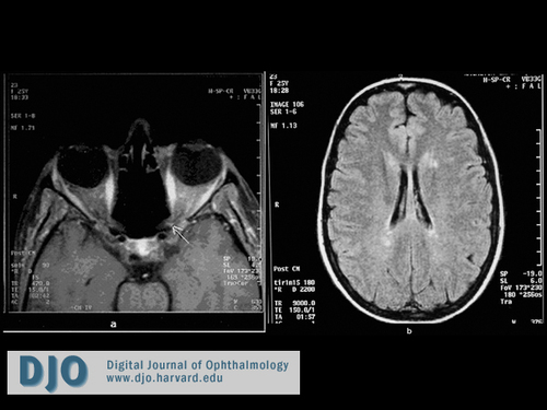
The patient also underwent magnetic resonance imaging (MRI) of the brain and orbits, which revealed lesions consistent with demyelinating disease at the junction of the left optic nerve and optic chiasm. This is shown on T1-weighted, gadolinium-enhanced scans (Figure 3a). On T1-weighted scans, there were also multiple periventricular demyelinating lesions consistent with demyelinating disease (Figure 3b). | |
|
Figure 3
T1-weighted, gadolinium-enhanced MRI of brain and orbits. (a) Lesions consistent with demeylination plaques on the left side of the optic chiasm (arrow). (b) Lesions consistent with demyelinating areas in the periventricular region.
 |
|
|
|
|
|
|
|
 Welcome, please sign in


