|
|
 |
|
|
|
|
|
A 26-year-old man with renal failure and vision loss
Digital Journal of Ophthalmology 2008
Volume 14, Number 13
July 12, 2008
|
Printer Friendly
|
|
|
 Tarek Alasil
 | University of Southern California Mario Meallet | University of Southern California
|
|
|
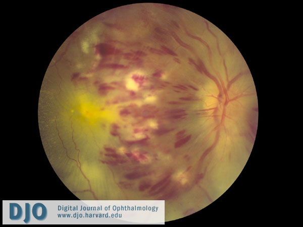
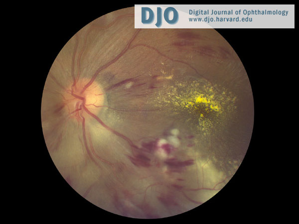
| Examination | | On ophthalmologic exam, the patient had a fixed, irregular, non-reactive right pupil that measured 4 mm. A right relative afferent pupillary defect (RAPD) was present. The left pupil was reactive to light. The patient had visual acuities of counting fingers on the right, and 20/200 on the left without improvement by pinhole. The intraocular pressure measured 12 mm Hg on the right and 10 mm Hg on the left. The slit-lamp exam was significant for a fixed pupil on the right and iris touching the cornea on the left. The fundus examination of the right eye revealed extensive intraretinal hemorrhages in all 4 quadrants, dilated and tortuous retinal veins, cotton wool spots, and disc edema (Figure 1). The fundus examination of the left eye revealed severe inferior disc swelling, intraretinal hemorrhages, and cotton-wool spots (Figure 2). | |
|
Figure 1
Color fundus photograph of the right eye shows extensive intraretinal hemorrhages in all 4 quadrants, dilated and tortuous retinal veins, cotton wool spots, and disc edema.
 |
|
|
Figure 2
Color fundus photograph of the left eye shows severe inferior disc swelling, intraretinal hemorrhages, and cotton-wool spots.
 |
|
|
|
|
|
|
|
 Welcome, please sign in