External examination demonstrated no abnormalities. Her visual acuity was assessed with the preferential looking test. With binocular viewing the result was 0.64 cpd (cycles per degree), corresponding to 20/960 Snellen acuity. For the right eye alone the visual acuity was 20/1,400 (Normal VA corrected for age is approximately 20/80). She was too restless for testing of the left eye. Her VEP (visual evoked potentials) acuities were 4.2 cpd on the right and 4.5 cpd on the left. These correspond to 20/143 and 20/133 Snellen acuity.
The pupils were round and equal and constricted in response to light. No afferent pupillary defect was seen. Additionally, using an infrared video system, paradoxical pupillary constriction to darkness was demonstrated. (see Video)
The cycloplegic retinoscopy was +5.50 sphere in both eyes. She was orthotropic on muscle balance testing. She had full ductions and versions. There was a small amplitude, high frequency, horizontal nystagmus. The direction of nystagmus did not change in different directions of gaze. There was no anomalous head posture and the patient did not appear to have a null point in which the nystagmus dampened.
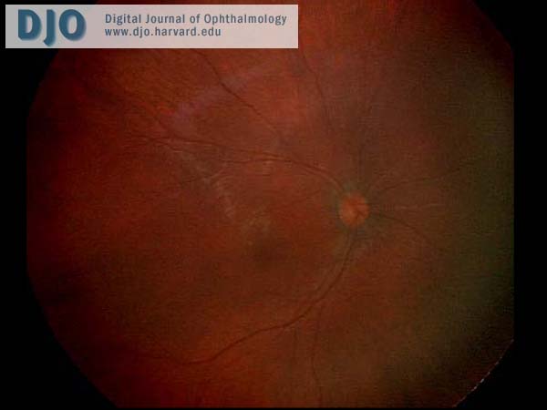
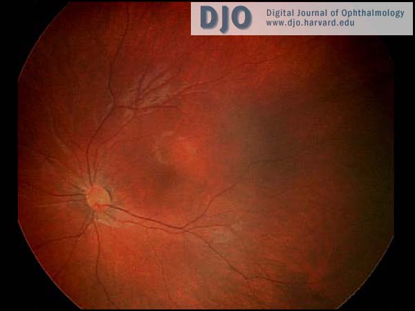
The anterior segment examination was normal. There were no iris transillumination defects. The intraocular pressures were normal at 8 mmHg in both eyes. On visual field testing, she looked for the hand light in all quadrants with both eyes. On indirect ophthalmoscopy, the media were clear; the optic nerve heads were pink with sharp margins and were normal in diameter. In the maculae, foveal reflexes were identifiable. The retinal vasculature appeared to be of normal caliber and distribution and no abnormalities were seen in the extramacular fundi. (see Figures 1 & 2)
 Welcome, please sign in