|
|
 |
|
|
|
|
|
A 36-year-old man with a red eye
Digital Journal of Ophthalmology 2008
Volume 14, Number 9
April 13, 2008
|
Printer Friendly
|
|
|
|
|
|
|
| Examination | On examination, the visual acuity was 20/20-2 in the right eye. The right pupil was reactive (6 mm to 3 mm) and the intraocular pressure was 14 mm Hg by applanation. His right eye extraocular movements were intact, and his visual field was full to confrontation. He had a prosthesis over his left eye.
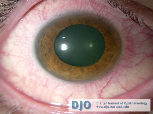
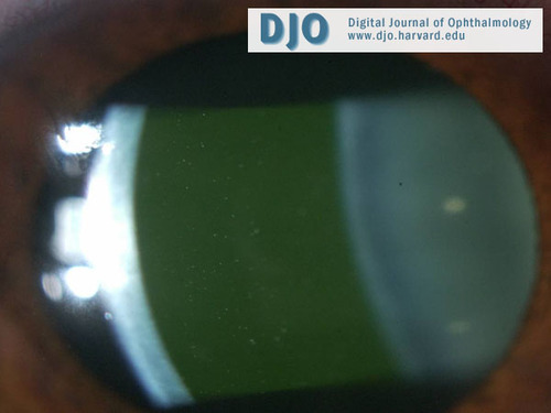
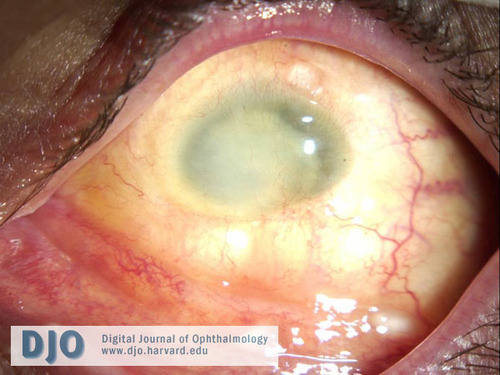
Slit lamp examination (see figures 1-3) revealed a scleral shell prosthesis on the left. His eyelids were flat without erythema or swelling bilaterally. His conjunctiva had 3+ injection with ciliary flush on the right and was quiet on the left. There were fine keratic precipitates on the right cornea. The left cornea was opaque due to pannus. There was no view of any deeper structures in the left eye. The right eye had 4+ cell and flare in the anterior chamber, a round and regular iris with no synechiae, and a clear lens. There was vitreous haze in the right eye.
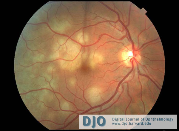
Funduscopic exam of the right eye (see figure 4) showed a normal optic nerve with a cup to disc ratio of 0.3. The vessels were within normal limits, but there were multiple creamy, placoid, deep retinal lesions extending from the right macula to the temporal periphery. There was no view of the left fundus. | |
|
Figure 1
Photograph of the right eye (post-dilation) displaying ciliary flush.
 |
|
|
Figure 2
Photograph of the right eye demonstrating anterior chamber cell and flare.
 |
|
|
Figure 3
The phthisical left eye after the scleral shell was removed showed no conjunctival injection. According to the patient, he was shot in the left eye 22 years ago. The eye was not removed as there was hope that he would regain vision. However 3 years later when the eye had become phthisical, he was fitted with a scleral shell.
 |
|
|
Figure 4
Fundus photo of the right eye demonstrating multiple creamy, placoid, deep retinal lesions in the macula and temporal periphery. The optic nerve and vessels were normal.
 |
|
|
|
|
|
|
|
 Welcome, please sign in