|
|
A 36-year-old man with a red eye
Digital Journal of Ophthalmology 2008
Volume 14, Number 9
April 13, 2008
|
Printer Friendly
|
|
|
|
|
|
|
| Ancillary Testing |
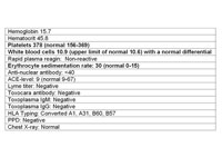
| A fluorescein angiogram was performed (see Figures 5-8). Laboratory studies and chest x-ray results are listed in Table 1. |
|
|
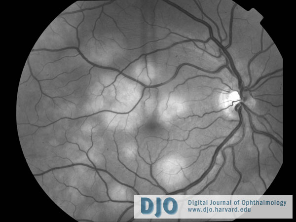
Figure 5
A red free photo showing placoid lesions of the right fundus.
 |
|
|
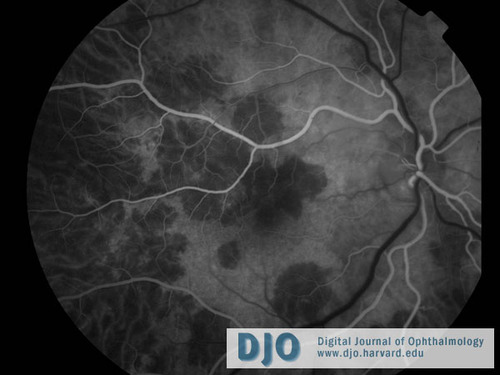
Figure 6
The arterial phase of the right eye fluorescein angiogram shows early blockage of the lesions.
 |
|
|
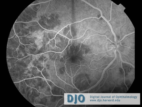
Figure 7
Venous phase of the right eye fluorescein angiogram.
 |
|
|
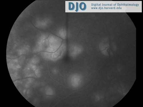
Figure 8
The late phase of the right eye fluorescein angiogram demonstrates late hyperfluorescence of the lesions.
 |
|
|
Table 1
Labs and studies that were obtained are listed above with abnormal values in bold.
 |
|
 Welcome, please sign in