|
|
 |
|
|
|
|
|
A 39-year-old man with painful proptosis after dental extraction
Digital Journal of Ophthalmology 2008
Volume 14, Number 4
February 7, 2008
|
Printer Friendly
|
|
|
 Brett Kotlus
 | Allure Medical Spa Veena Kumar | Maxwell Aesthetic Surgery Robert Cravens | Tucson Ear Nose and Throat Robert Dryden | Arizona Centre Plastic Surgery
|
|
|
| Ancillary Testing | Radiographic Studies
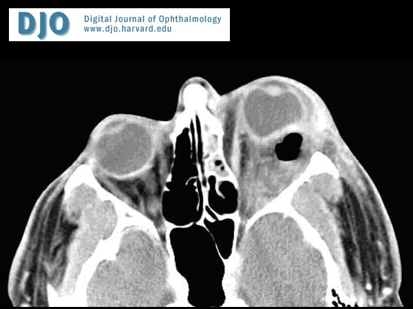
Computed tomography of the head was performed, revealing opacification of the left anterior ethmoid and maxillary sinuses. In addition, an intraconal collection of fluid with an adjacent gas bubble was identified lateral to the optic nerve, exerting mass effect on the posterolateral aspect of the globe. | |
|
Figure 2
Axial CT image demonstrating intraconal gas with posterolateral globe deformation.
 |
|
|
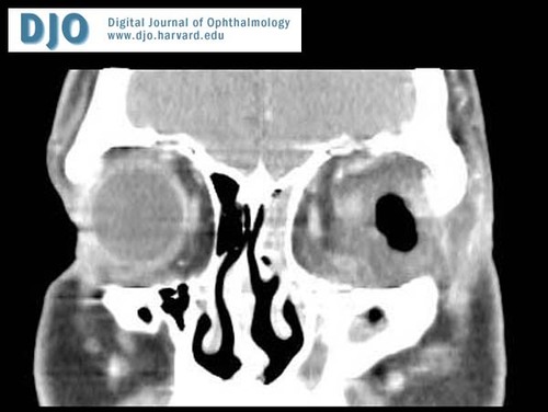
Figure 3
Coronal CT image
 |
|
|
|
|
|
 Welcome, please sign in