Welcome, please sign in
Follow DJO on Facebook Follow DJO on Twitter
Grand Rounds
  Most Recent Cases
  Dates of Case
  Type of Case
  Submit a Grand Round.
  Register with DJO to receive personalized updates.

If you're already a
member, please sign in.
A 38-year-old man with bilateral foveal hemorrhages
Digital Journal of Ophthalmology 2008
Volume 14, Number 7
March 11, 2008
Printer Friendly


Ian Yeung | Southampton General Hospital, UK
Conor Mulholland | The Royal Victoria Hospital, Belfast
Examination
Visual acuities without correction were 6/18 (20/60) OD and 6/24 (20/80) OS with no improvement with pinhole.

The anterior segment examination was normal.

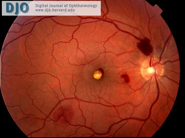
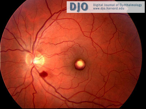
Dilated fundoscopic examination showed bilateral foveal preretinal hemorrhages and bilateral scattered nerve fiber layer hemorrhages. The foveal hemorrhages were beginning to organize at the time of presentation to our department.

Blood pressure, blood glucose and complete blood count were normal. Specifically, there was no evidence of anemia.
top
Right eye
Fundus photograph demonstrating foveal pre-retinal hemorrhage and scattered nerve fiber layer hemorrhages.

Left eye
Fundus photograph demonstrating organizing foveal pre-retinal hemorrhage and a nerve fiber layer hemorrhage in the inferior arcade.