Orbit/Oculoplastics Quiz 17
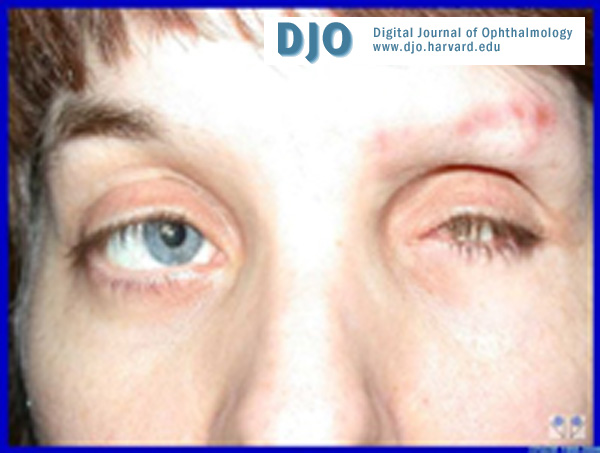
Clinical photo
There is significant enophthalmos OS due to the shrunken globe. The patient is mentally retarded and has been noted to pick at her left eyebrow because of the pain she's been having in that eye.
There is significant enophthalmos OS due to the shrunken globe. The patient is mentally retarded and has been noted to pick at her left eyebrow because of the pain she's been having in that eye.

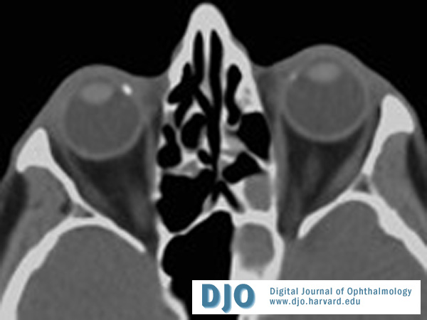
CT scan of the patient
There is significant atrophy and disorganization OS. Within the eye, there is dense calcification which can often be seen in phthisical eyes.
There is significant atrophy and disorganization OS. Within the eye, there is dense calcification which can often be seen in phthisical eyes.
Senile calcific plaques on CT
There is a focal hyperdensity seen nasally OU that correspond to the typical location of senile calcific plaques.
There is a focal hyperdensity seen nasally OU that correspond to the typical location of senile calcific plaques.
Clinical appearance of senile calcific plaques
There is a slight elevation of the sclera seen temporally. This can be both temporal and nasal, and can appear bilaterally.
There is a slight elevation of the sclera seen temporally. This can be both temporal and nasal, and can appear bilaterally.
Answer: Phthisis bulbi is the end-stage ocular response to injury or severe disease of the eye. Clinically, it is characterized by a soft atrophic eye with disorganization of intraocular structures. Phthisical eyes can sometimes be painful and may require medical or surgical treatment.
2. What is the etiology of this condition?
Answer: Ocular injury, radiation, infection, or diffuse disease can all lead to phthisis bulbi. Initial damage to intraocular structures either from penetrating trauma or inflammation can eventually lead to widespread atrophy and disorganization of the eye. In the case of retinoblastoma, phthisis can occur as a consequence of tumor necrosis and subsequent inflammation in the eye.
3. What is the natural history of this condition?
Answer: Atrophy of intraocular contents, including the retina and uvea, can initially occur without any evidence of shrinkage. Over time, atrophy of the globe can lead to hypotony and shrinkage while maintaining recognizable structures within the eye (atrophia bulbi). When atrophy and scarring of the intraocular structures leads to disorganization, the term phthisis bulbi is used.
4. What are the clinical and radiologic characteristics of this condition
Answer: A phthisical eye can grossly have the characteristic shrunken appearance that may be cuboidal in shape secondary to the pull of the rectus muscles on the soft globe. Retinal and ciliochoroidal detachments occur with hypotony, and the sclera can be markedly thickened. Even on CT scan, intraocular structures are not easily identifiable and there is often calcification seen within the eye. In phthisis bulbi, intraocular calcium may be deposited within band keratopathy, a cataractous lens, bony metaplasia of the RPE, sclera, a gliotic neural retina, or optic nerve.
5. What is the differential diagnosis for intraocular calcification?
Answer: Tumors (notably, retinoblastoma and bony tumors such as osteoma) are another important cause of intraocular calcification and should be in the differential when assessing a blind painful eye. In one series, 2.7% of patients with retinoblastoma had concomitant phthisis bulbi. There are also reports of phthisical eyes found to have malignant tumors such as osteogenic sarcoma and uveal melanoma. For these reasons, it is important to send enucleated phthisical eyes for histopathology.
Ocular conditions other than tumors that present with intraocular calcium includes band keratopathy, senile calcific plaques, dystrophic calcification associated with infection, intraocular foreign body or implants, and choroidal calcifications.