Orbit/Oculoplastics Quiz 13
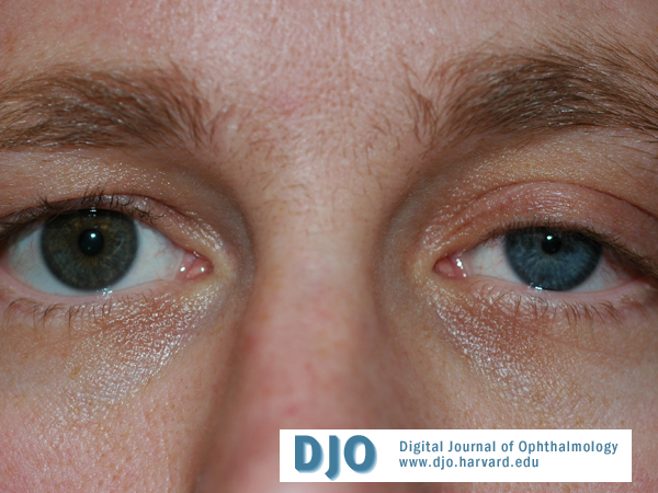
Figure 1
Answer: The right pupil will dilate in response to both. The left pupil will not dilate after cocaine but will do so after hydroxyamphetamine.
Horner’s syndrome is characterized by anisocoria greater in low light. The pupil with loss of sympathetic innervation dilates poorly. In all types of Horner’s the affected pupil dilates less than the unaffected pupil in response to cocaine. Pupillary response to hydroxyamphetamine is used to differentiate a third from a first or second order neuron abnormality. Failure to dilate after hydroxyamphetamine indicates a third order abnormality.
2. 2. Why might he hold a near card closer to his left eye?
Answer: Some patients with Horner’s syndrome demonstrate enhanced accommodation in the affected eye.
There are many signs that may be associated with Horner’s syndrome, due to loss of sympathetic innervation. These include ipsilateral upper and lower lid ptosis, the combination of which may cause pseudo-enophthalmos. Conjunctival congestion, miosis, dilation lag, decreased intraocular pressure, and increase in accommodation with normal light and pupillary responses. In third order Horner’s anhidrosis occurs on the ipsilateral medial forehead and nose. In cases in which the lesion is proximal to the carotid bifurcation, anhidrosis occurs on the entire ipsilateral hemi-face. First order lesions cause increase in ipsilateral hemi-body sweating. Level of perspiration may be tested by application of alizarin powder, which darkens on contact with sweat. Iris heterochromia is usually associated with congenital Horner’s. There are case reports of iris heterochromia in acquired disease. (1) There is also a report of neurotrophic endothelial failure associated with Horner’s. (2)
1. Diesenhouse MC Palay DA Newman NJ Acquired heterochromia with Horner’s syndrome in two adults, Ophthal 1992; 99:1815-17.
2. Zamir E Chowers I Banim E Neurotrophic corneal endothelial failure complicating acute Horner’s syndrome, Ophthalm 1999;106:1692-6.
3. 3. If the onset of these symptoms and signs were acute, what workup would be appropriate?
Answer: 1. History: Examination of old photographs to determine duration of symptoms. Occurrence of headache or arm pain, history of stroke, surgery or trauma involving the head, neck, or upper chest. Heavy smoking.
2. Physical exam: Supraclavicular nodes, thyroid enlargement, neck masses, scars.
3. Determine order of neuron involved with cocaine/hydroxyamphetamine
First order lesions:
Descending sympathetic chain hypothalamus, brainstem, or spinal cord C8-T2. The most common cause is Wallenberg syndrome, or lateral medullary stroke, which is caused by infarction of the posterior inferior cerebellar or lateral medullary artery. Associated symptoms include but are not limited to skew deviation, lateropulsion, poor smooth pursuit, nystagmus, ataxia, ipsilateral hemi-facial and contralateral hemi-body anesthesia, vertigo, nausea, vomiting, dyspohagia, vocal cord paralysis. Other common causes are trauma or surgery involving C8-T12, syringomyelia, myelitis, tumors, multiple sclerosis, and Brown-Sequard syndrome.
Second order lesions:
The most common cause is a compressive or traumatic thoracic/neck injury. Injuries include brachial plexus trauma, neck tumors, apical lung tumors, lung, mediastinal or neck surgery, chest tube or arterial or venous line migration, Swan-Ganz catheterization, coronary artery bypass grafting. Of note, Pancoast tumors may cause increased sympathetic stimulation and ipsilateral facial flushing and hyperhydrosis prior to causing Horner’s.
Third order lesions:
Ipsilateral head pain associated with Horner’s is called Raeder's paratrigeminal syndrome or neuralgia. Ominous disorders associated with Raeders include carotid artery dissection and thrombosis and cavernous sinus lesions. Carotid dissection or thrombosis usually follows trauma and also may be associated with connective tissue disease. Associated symptoms include subjective bruit, carotidynia, dysgeusia, and signs of cerebral ischemia. HORNERS SYNDROME IS THE MOST COMMON NEUROLOGIC SIGN OF CAROTID DISSECTION, OCURRING IN 50% OF PATIENTS. (3,4) Painful Horner’s is carotid dissection until proven otherwise. Cavernous sinus and orbital apex syndromes present with associated dysfunction of cranial nerve III, IV, and VI. In a child, neuroblastoma and birth trauma must be considered.
Less ominous causes of painful third order Horner’s are cluster headache and small vessel ischemic disease, including diabetes. Oral surgery or trauma may injure the superior cervical ganglion or internal carotid artery at the peritonsillar area. Autonomic neuropathies such as amyloid or diabetes may also cause Horner’s.
Work-up includes appropriate studies including neurologic exam, examination of the oral cavity, MRI/MRA of ICA, neck, cavernous sinus, or brain, chest or abdominal CT.
3. Hart RG, Easton JD, Dissection of cervical and cerebral arteries. Neurol Clin, 1983;1:155-82.
4. Mokri B, Sundt TM, Houser OW, et al, spontaneous dissection of the cervical internal carotid artery, Ann Neurol. 1986;19:126-138.
4. 4. Considering the results of the test pictured above, what is the most efficient surgical intervention?
Answer: Mullerectomy may be considered when there is improvement in ptosis in response to phenylephrine application. If the lid fails to elevate with phenylephrine application, levator advancement would be required.