Orbit/Oculoplastics Quiz 9

Figure 1
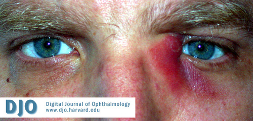
42-year-old man with a seven-day history of erythema and tenderness near the left medial canthus.
42-year-old man with a seven-day history of erythema and tenderness near the left medial canthus.

Figure 2
Patient with acute dacryocystitis who received medical management.
Patient with acute dacryocystitis who received medical management.
Answer: The photograph shows acute dacryocystitis, or inflammation of the tear sac. Tenderness and erythema evolve over a period of days. Epiphora is almost always present, and disturbance of the tear film may reduce vision. Purulent punctal discharge is often noted, particularly if pressure is applied to the lacrimal sac. However, this does not always occur as the valve of Rosenmueller may prevent reflux. Fever and prostration may occur. The condition is distinct from chronic dacryocystitis, which is characterized by epiphora and less fulminant inflammation. In children it should be remembered that enceophalocele/meningomyeloceles are in the differential of medial canthal masses.
2. What is the underlying cause of this condition?
Answer: Nasolacrimal duct obstruction causes tears to stagnate in the lacrimal sac, which in turn fosters infection. Causes of acquired nasolacrimal duct obstruction (or stenosis) include inflammatory disease, facial trauma, tumor, or other nasal or ethmoid disease. However, most cases are idiopathic. The patient shown in Figure 1 has sarcoidosis.
3. What organisms are usually involved?
Answer: Gram-positive flora of the conjunctiva, such as Staphylococcus and Streptococcus, are usually responsible. E. coli, Pseudomonas, other Gram-negative organisms, and fungi are involved in a minority of cases. In patients with a history of chronic sinus disease, immunocompromised status, dental infection, prior major facial sutrgery, etc., the antibiotics may be broadened to include anerobic and gram negative bacteria.
4. What are the possible complications of this condition?
Answer: Preseptal cellulitis and conjunctivitis are common complications. Cutaneous fistulas may form from the lacrimal sac. Less common but more severe complications include orbital cellulitis and sepsis.
5. What is the appropriate treatment?
Answer: All patients benefit from warm compresses, lacrimal sac massage, and analgesics. Most cases merit oral antibiotics such as amoxicillin-clavulanate. Computed tomography is helpful for evaluating the cause of nasolacrimal duct obstruction when history is suggestive of a possible defined cause (such as prior trauma). Children and anybody with suspected orbital cellulitis should be admitted for intravenous antibiotics. A pointing or non-healing abscess (such as that in Figure 1) should be incised, drained, and allowed to heal by secondary intention (in contrast, the patient shown in Figure 2 received medical management). When surgical treatment is necessary, it is important to note that two distinct collections may be present, one immediately beneath the skin and a second within lacrimal the sac itself. Both must be drained in order to address the infection. Lacrimal system probing is not indicated during the acute infection. After the acute infection has resolved, dacryocystorhinostomy (DCR) is performed to correct the nasolacrimal duct obstruction and prevent future dacryocystitis. In older patients with a history of dry eye no prior tearing, and who might be poor surgical candidates, dacryocystectomy may be considered as an alternative to DCR. In young patients with aquired (not congenital) NLDO, a search for an etiology must be performed, including nasal examination, possible CT scan, and biopsy of the lacrimal sac at the time of DCR.