Retina/Uveitis Quiz 16
Rosa Y Kim, MD | Massachusetts Eye and Ear Infirmary, Harvard Medical School
February 12, 1997
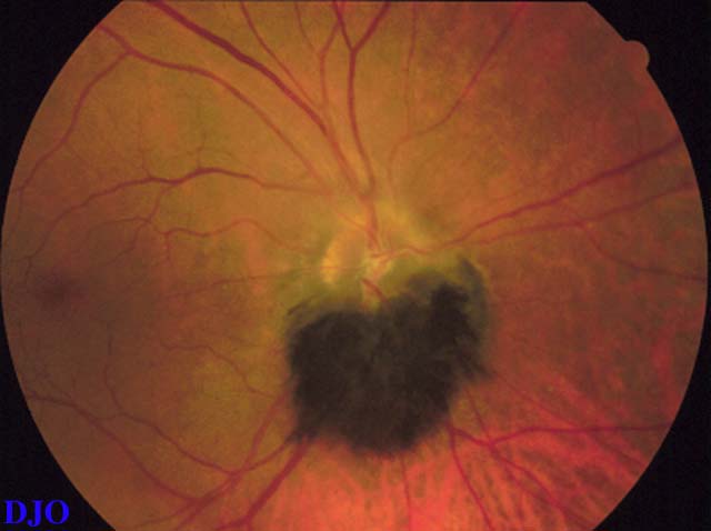
Figure 1
A darkly pigmented, elevated, lesion at the optic nerve head OD in an asymptomatic patient.
A darkly pigmented, elevated, lesion at the optic nerve head OD in an asymptomatic patient.
Answer: Melanocytoma, RPE hyperplasia, congenital hypertrophy of the RPE, combined hamartoma of the RPE and sensory retina, and melanoma
2. What is the most likely diagnosis?
Answer: Melanocytoma of the optic nerve head
3. Is this lesion malignant?
Answer: Melanocytomas are benign, highly pigmented tumors arising FROM melanocytes. They are a variant of nevi. However, malignant transformation of these lesions has been reported and confirmed histopathologically.
4. Is there any sex or race predilection?
Answer: Both sexes are equally affected. The incidence is increased in blacks. Interestingly, melanomas are extremely rare in this group.
5. What is the most common visual field defect in patients with this lesion?
Answer: An enlarged blind spot is the only visual disturbance. Except for large tumors, these lesions are asymptomatic and dignosed during routine fundus examination.