Orbit/Oculoplastic Quiz 1: Orbit

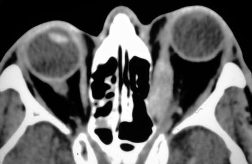
Figure 1
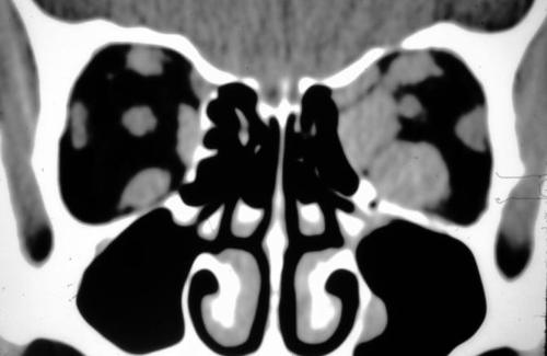
Figures 1-2. This is the orbital CT of a man with painless progressive proptosis OS
Figures 1-2. This is the orbital CT of a man with painless progressive proptosis OS

Figure 2
Answer: Thyroid ophthalmopathy. This is the most common cause of unilateral as well as bilateral proptosis. The absence of pain and the enlargement of muscles without involvement of the tendinous portion of the muscle is typical of thyroid disease.
2. What is the differential diagnosis of enlarged extraocular muscles?
Answer: Thyroid disease is the most common cause of enlarged extraocular muscles. Usually, the degree of asymmetry between the orbits is not as pronouced as in this patient. Inflammatory orbital pseudotumor can cause enlargement of the muscle, but the tendinous portion is classically affected by the inflammation, in contrast to thyroid disease where the tendinous portion is spared. Rarely, neoplastic lesions may infiltrate the muscles. Amyloidosis may also enlarge the muscles through an infiltrative process.
3. Which extraocular muscles are mont commonly involved in this disorder?
Answer: The extraocular muscles which are affected, FROM most to least are the: inferior rectus, medial rectus, superior rectus, and lateral rectus.
4. What are potentially sight threatening complications in this disorder?
Answer: Compressive optic neuropathy FROM muscle enlargement and corneal exposure leading to ulceration and perforation.
5. How would you manage this condition?
Answer: Treatment is dependent on the severity of the disease. If mild, the patient may be treated with topical lubrication and patching at night. If in the acute inflammatory phase, the patient may require systemic corticosteroids or immunosuppressive agents. Lid surgery may be necessary to REPAIR lid retraction. Orbital decompressive surgery is an effective treatment for compressive optic neuropathy. Traditionally this was done by removing the medial orbital wall along with the orbital floor. However some advocate to remove the medial and lateral wall while leaving the orbital floor intact. The combined medial and lateral wall decompression is usually sufficient to achieve the surgical goals. The medial wall decompression can be performed endoscopically. The disadvantages of floor decompression includes the inability to predict the results. The patient may also experience enophthalmos due to excessive herniation INTO the maxillary sinus.