Neuro-ophthalmology Quiz 7
Howard Pomeranz, M.D., Ph.D | Massachusetts Eye and Ear Infirmary, Harvard Medical School, University of Maryland Medical Center
M.A. Afshari, M.D., M.P.H. | Massachusetts Eye and Ear Infirmary, Harvard Medical School
August 3, 1997
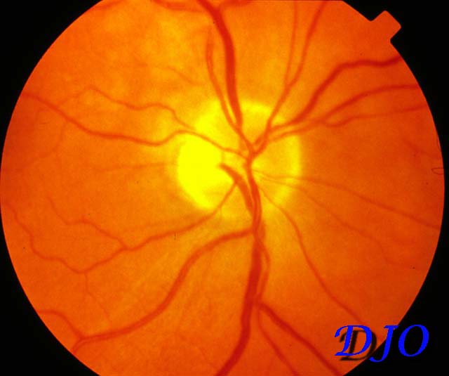
Figure 1
Figures 1-2. Fundus Photographs
Figures 1-2. Fundus Photographs
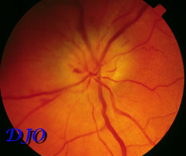
Figure 2
Answer: The photographs demonstrate a normal appearing right optic nerve with a small cup to disk ratio. The left optic nerve is edematous with several flame hemorrhages in the nerve fiber layer and on the optic nerve head. The ophthalmic history and findings are suggestive of nonarteritic anterior ischemic optic neuropathy.