Neuro-ophthalmology Quiz 1
Yichieh Shiuey, MD | Massachusetts Eye and Ear Infirmary, Harvard Medical School
July 15, 1996
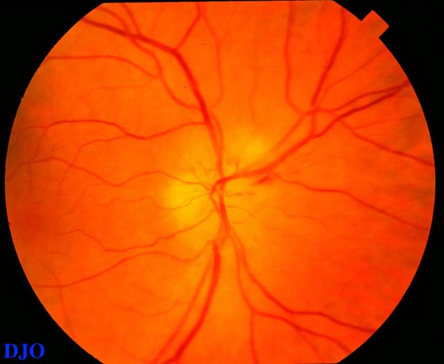
Figure 1
This is the view of the right optic disc of a 73 year old woman with a sudden decrease in vision of the right eye. The patient had a history of headache for several months and also complained of joint pains and anorexia. The fundus of the left eye was normal
This is the view of the right optic disc of a 73 year old woman with a sudden decrease in vision of the right eye. The patient had a history of headache for several months and also complained of joint pains and anorexia. The fundus of the left eye was normal
Answer: Arteritic and non-arteritic anterior ischemic optic neuropathy (AION) as well as bulbar optic neuritis are in the differential. Because of the patient's systemic symptoms and the pale quality of the disc edema, arteritic anterior ischemic optic neuropathy is the most likely diagnosis.
2. What kind of laboratory workup does this patient need?
Answer: Laboratory studies to rule out temporal arteritis include an erythrocyte sedimentation rate (ESR) and a complete blood count (CBC). Most patients with temporal arteritis will have an elevated ESR. The normal upper LIMIT for the ESR in men is the patient's age divided by two. For women the normal upper LIMIT for the ESR is the patient's age divided by two, plus ten. A CBC is useful to exclude other systemic diseases that may cause an optic neuropathy e.g. leukemia.
3. If this patient had an elevated erythrocyte sedimentation rate which diagnostic procedure would you perform next?
Answer: A history consistent with temporal arteritis associated with an elevated ESR, requires the ophthalmologist to obtain a temporal artery biopsy. If the biopsy is positive the diagnosis is confirmed. However, a normal temporal artery biopsy does not exclude the diagnosis of temporal arteritis. A false negative rate of 5% to 9% has been reported in the literature. If the clinical suspicion is high and the first biopsy is normal, the opposite temporal artery should be biopsied.
4. In patients with temporal arteritis affecting one eye, what percentage of patients will go on to have involvement of the other eye? If there is involvement of the other eye, when does this usually occur?
Answer: One study showed that more than one third of patients who suffered vision loss prior to treatment of one eye developed bilateral involement. Typically, the second eye becomes involved within days or weeks of the first eye in arteritic AION. This is in contrast to non-arteritic AION where the second eye becomes involved in months to years.
5. How would you treat temporal arteritis?
Answer: Corticosteroids are indicated for the treatment of temporal arteritis. However, there are no controlled clinical trials which have addressed the optimal dosage or duration of corticosteroid in this condition. There is evidence in the literature that initial daily doses of 50 to 75 mg per day of oral prednisone is effective in preventing vision loss in patients that have not already lost vision. High dose intravenous steroids (e.g. 1000 mg of IV methylprednisolone q 12 hours for 5 days) have been advocated by some for patients which have already lost vision in one eye, in ORDER to prevent bilateral blindness.