Cornea/Refractive Surgery Quiz 20
Yichieh Shiuey, MD | Massachusetts Eye and Ear Infirmary, Harvard Medical School
February 9, 1998
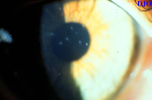
Figure 1
Figures 1-2. These are the anterior segment photographs of the right eye of a patient with recurrent episodes of foreign body sensation and tearing OU. The left eye has a similar appearance on slit lamp examination.
Figures 1-2. These are the anterior segment photographs of the right eye of a patient with recurrent episodes of foreign body sensation and tearing OU. The left eye has a similar appearance on slit lamp examination.
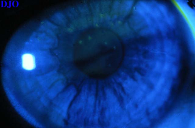
Figure 2