Cornea/Refractive Surgery Quiz 15
Scott Burk, MD, PhD | Massachusetts Eye and Ear Infirmary, Harvard Medical School
January 21, 1997
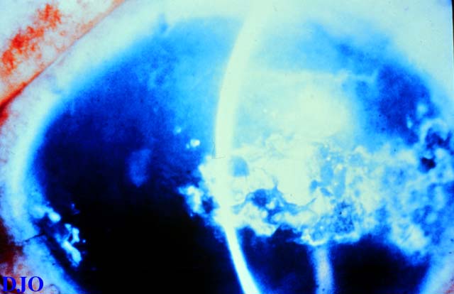
Figure 1
Figures 1-3. Anterior segment photographs of 3 different individuals.
Figures 1-3. Anterior segment photographs of 3 different individuals.

Figure 2
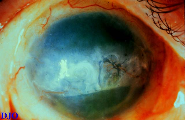
Figure 3
Answer: Band keratopathy.
2. What is the pathophysiology of this condition?
Answer: Deposition of calcium (hydroxyapatite) in the epithelial basement mermbrane, Bowman's layer, and superficial stroma. Generally the factors leading to calcium deposition are the concentration of calcium in the tear film and the soluability, which is related to the phosphorus concentration and the pH.
3. What underlying conditions may result in this finding?
Answer: (1) chronic ocular disease such as , uveitis, interstitial keratitis, and phthisis, (2) hypercalcemia of any cause, (3) hyperphosphatemia, (4) hereditary band keratopathy, (5) toxic and mercurial vapors, (6) silicone oil in aphakic eyes.
4. What is the usual presentation for this condition?
Answer: The disease usually begins at the interpalpebral limbus as a fine white granularity and gradually spreads over the interpalpebral corneal surface.
5. What treatment would you recommend?
Answer: Calcium deposits can usually be removed by superficial keratectomy with or without chelating the calcium with EDTA. Excimer phototherapuetic keratectomy is also a good choice for treatment. Treating the underlying condition of course is beneficial.
6. Does this condition recur?
Answer: Yes, Band keratopathy may slowly recur. Treatment may then be repeated.