A 51-year-old man with photopsias
Digital Journal of Ophthalmology 2008
Volume 14, Number 8
April 4, 2008
Volume 14, Number 8
April 4, 2008
Saqib Ali Khan Utman | Furness General Hospital, Barrow In Furness, UK
Benjamin J. R. Moate | Furness General Hospital, Barrow, UK
The right inferotemporal retina (Figures 1 and 2) excluding the macula had gross pigmentary changes. At the 7 o’clock position in the periphery, a small nodule of neovascularization was seen. These features were consistent with spontaneous reattachment of the retina. There was a slight swelling of apparently atypical retinoschisis in the superotemporal periphery from 10 to 12 o’clock. Yellow-white vitreous opacities were also seen.
Examination of the left fundus (Figure 3 and 4) revealed localized inferotemporal dialysis surrounded by minimal subretinal fluid and a well-established demarcation line.
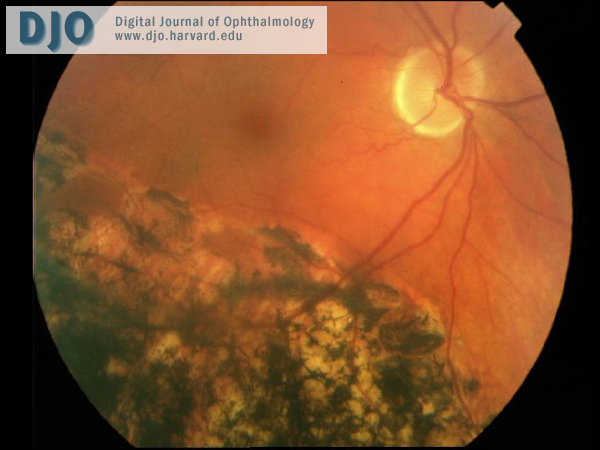
Figure 1
Photograph of the right fundus
Photograph of the right fundus
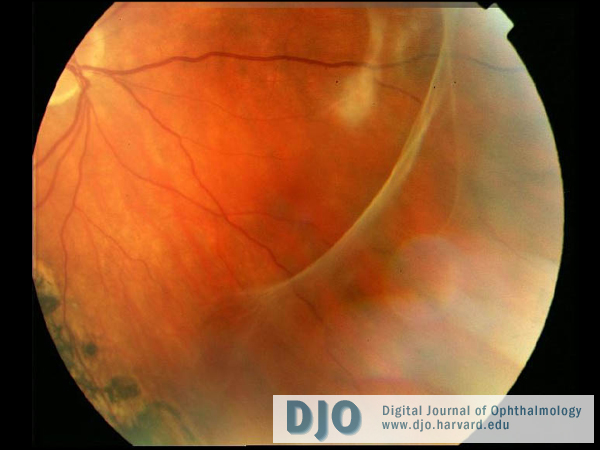
Figure 2
Photograph of the right fundus
Photograph of the right fundus
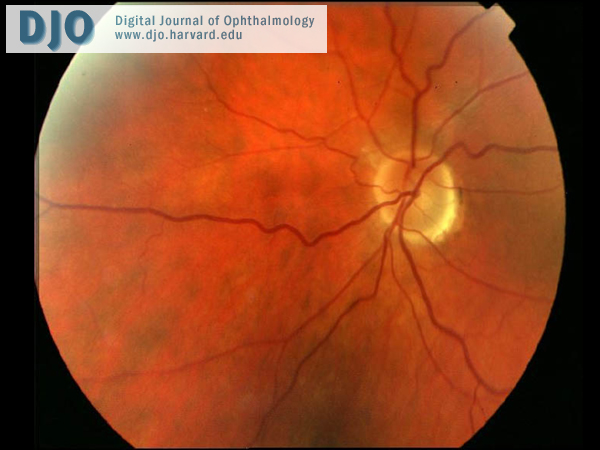
Figure 3
Photograph of the left fundus
Photograph of the left fundus
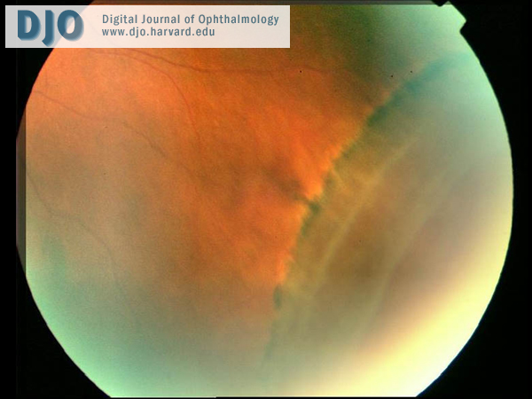
Figure 4
Photograph of the left fundus
Photograph of the left fundus
In old retinal detachments, retinal thinning secondary to atrophy, secondary intraretinal cysts, subretinal demarcation lines and retinal holes or tears are seen.
Degenerative Retinoschisis
Degenerative retinoschisis is a smooth, convex, thin and immobile elevation of layers of retina. However, demarcation lines and secondary cysts in the inner leaf are usually absent in retinoschisis.
Uveal Effusion syndrome
The uveal effusion syndrome is a rare, idiopathic condition characterized by choroidal detachment associated with exudative retinal detachment.
Retinal dialysis is probably secondary to a developmental abnormality of the inferotemporal periphery of the retina and vitreous base and may be precipitated by trauma.(2) In some cases, an autosomal recessive mode of inheritance (3) and the possible involvement of lattice degeneration have been considered.(4)
Inferotemporal quadrant retinal dialysis occurs in young adults and is associated with demarcation lines, retinal cysts, peripheral microcystoid degeneration and yellow-white vitreous opacities. The majority of these features were seen in our patient.
Genetic factors play a role in some cases of inferotemporal dialysis, the cause of which is probably multifactorial.(5)
2. Kinyoun JL, Knobloch WH. Idiopathic retinal dialysis. Retina. 1984 Winter-Spring; 4(1):9-14
3. Verdaguer TJ, Rojas B, Lechuga M. Genetical studies in nontraumatic retinal dialysis. Mod Probl Ophthalmol. 1975; 15:34-9.
4. Smiddy WE, Green WR. Retinal dialysis: pathology and pathogenesis. Retina. 1982; 2:94-116.
5. Vaiser A, Jost BF. Bilateral inferotemporal dialysis in identical twins. Ann Ophthalmol.1992 Oct; 24(10):378-80.