A 33 year old man with difficulty adapting to light
Digital Journal of Ophthalmology 2004
Volume 10, Number 6
April 24, 2004
Volume 10, Number 6
April 24, 2004
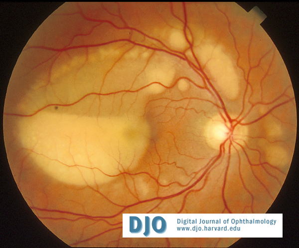
Figure 1
Numerous exudative retinal detachments with yellowish intra and subretinal depositions
Numerous exudative retinal detachments with yellowish intra and subretinal depositions
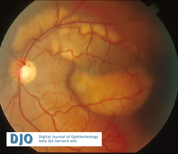
Figure 2
Numerous exudative retinal detachments with yellowish intra and subretinal depositions
Numerous exudative retinal detachments with yellowish intra and subretinal depositions
Fluorescein angiography (figure 3) demonstrated hypofluorescence of the lesions due to blockage of dye transmission by the yellow deposition material.
A scotopic and photopic full-field ERG were normal. An EOG was markedly depressed (Arden ratio of 1.3 in both eyes ).
The results of a complete blood workup were negative except for elevated hepatic enzymes. Further investigations led to the diagnosis of hepatitis C. No risk factors were established.
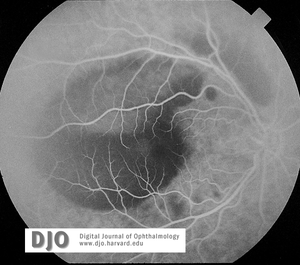
Figure 3
Hypofluorescence due to blockage of dye transmission by the yellow deposition material
Hypofluorescence due to blockage of dye transmission by the yellow deposition material
Atypical central serous chorioretinopathy
Posterior scleritis
Uveal effusion syndrome
Sarcoidosis and other inflammatory diseases
Multifocal Best’s disease is an unusual form of this disorder that can occur in patients without family history(4,5,6). The reduction in visual acuity seems more rapid and severe and the lesions can appear, disappear or coalesce(4).
In this case, the additional presence of hepatitis C (without any known risk factors) is an atypical feature. An extensive review of the english literature did not reveal any known association between hepatitis C and Best’s disease.
2)Weingeist T, Kobrin J, Watzke R. Histopathology of Best’s macular dystrophy. Arch Ophthalmol 1982 ; 100 : 1108-1114
3)Mohler CW, Fine S. Long-term evaluation of patients with Best’s vitelliform dystrophy. Ophthalmology 1981 : 88 : 688
4) Caumon C,Bernard JA, Mondon H, Quentl G, Hamard H, Chevaleraud J. Multifocal vitelliform dystrophy : a case report. Journal Français d’ophtalmologie 1981 : 4 (4) : 279-85
5) Miller S. Multifocal Best’s vitelliform dystrophy. Arch Ophthalmol 1977 : 95 : 984-90
6) Ciulla TA, Frederick AR Jr. Acute progressive multifocal Best’s disease in a 61-year-old man. Am J Ophthalmol 1997 : 123 (1) : 129-31