20 year old man with eyelid swelling
Digital Journal of Ophthalmology 2003
Volume 9, Number 3
October 31, 2003
Volume 9, Number 3
October 31, 2003
Previous ocular history: None
Past medical history: None
Pupils: Normal OU
Motility: Full OU
Tonometry: Normal OU
Slit lamp examination of the anterior segment: Normal OU
Fundus examination: Normal OU
Eyelid exam: The RUL revealed a mild preseptal cellulitis around an indurated lesion which showed a central fistula draining a serosanguineous material. (Fig 1 and 2)
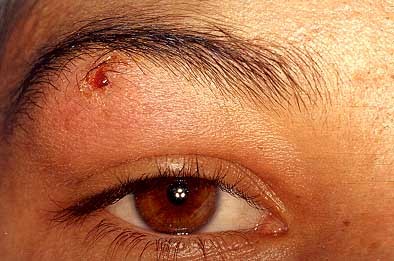
Figure 1
Eyelid swelling with central ulcerated lesion.
Eyelid swelling with central ulcerated lesion.
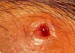
Figure 2
Higher magnification of ulcerated lesion.
Higher magnification of ulcerated lesion.
2. Pyogenic granuloma
3. Cystic lesion
4. Palpebral myiasis
Flies are arthropods of the class Insecta, some of which belong to the order Diptera with more than 80,000 species (1). Ophthalmomyiasis externa or palpebral myiasis, refers to the infestation of living land humans or vertebrates with certain dipterous larvae that feed on the host’s tissue (2). Although ophthalmomyiasis usually appears in the external tissues, a case of orbital infestation was reported by Kersten and co-authors (3). There are two basic classifications that grouped myiasis based either on the affected area, or according to its habits (4).
Ophthalmomyiasis has been associated with larvae from different species (Cutebra species, Oestrus ovis, Hypoderma bovis, and Dermatobia hominis). In Mexico as well as in central and south America, D. hominis is the most frequent. This big-sized fly has yellow head and legs, and purple thorax and abdomen. Infestation usually occurs in wooded areas and it is through an intermediary parasite. The adult fly never gets in contact with the host. The cycle starts when a female D. hominis captures an arthropod (usually a mosquito from the order Psorophora) (5) and places the eggs in the inferior part of its abdomen. When the mosquito charged with mature eggs is placed over the host, larvae are stimulated and penetrate the skin. Because of its aerobic characteristics, once the larva is in the host’s tissue, it cuts a hole through the skin in its search for oxygen, producing a furuncle-type lesion. These lesions are usually painful and may easily be over-infected.
Different treatment options have been reported. Some Indians from South America use fat or tobacco paste to close the hole and avoid oxygen entrance (1). Chloroform in olive oil and a 5% solution of carbolic acid have also been used to destroy the larvae (6). Many furunculous larvae may be simply expressed from the lesion. But in the case of D. hominis it is not possible, because it has mouth-hooks and body-hooks that firmly adhered to the skin. In these cases as happened with our patient, larvae need to be surgically removed.
It is very common that because of the furunculous appearance of the lesion, patients with external myiasis are previously treated with antibiotics and antiseptics. However, it is important to suspect infestation with Dermatobia hominis when itching and red swelling of the lid are present in patients who have been to endemic places in Central and South America (7).
Intra-operative video
Recovery of an alive insect larvae from the eyelid.
Click on image to begin video. Requires Windows Media Player which can be downloaded free from Microsoft by clicking here
Recovery of an alive insect larvae from the eyelid.
Click on image to begin video. Requires Windows Media Player which can be downloaded free from Microsoft by clicking here
2. Harwood RF, James MT. Entomology in human and animal health. 7th ed. New York: MacMillan, 1979: Chapter 13
3. Kersten RC, Shourkrey NM, Tabbara KF. Orbital myiasis. Ophthalmology 1986;93:1228-1232.
4. James MT. The flies that cause myiasis. Miscelaneous Publication. N°.631. Washington DC, US Dept of Agriculture, 1947:1-175.
5. Hubler WR, Rudolf AH, Dougherty EF: Dermal myiasis. Arch Dermatol 1974;110:109-110
6. Arenas R, Estrada R. Tropical dermatology. Landes Biosciences. Georgetown. 2001.
7. Bangsgaard R, Holst B, Krogh E, Heegaard S.Palpebral myiasis in a Danish traveler caused by the human bot-fly (Dermatobia hominis). Acta Ophthalmol Scand 2000 Aug;78(4):487-9