A 50-year-old man with a long-standing, large-angle exotropia and limitation of adduction in the left eye
Digital Journal of Ophthalmology 2013
Volume 19, Number 4
December 30, 2013
DOI: 10.5693/djo.03.2013.09.004
Volume 19, Number 4
December 30, 2013
DOI: 10.5693/djo.03.2013.09.004
Download PDF
Reshma A. Mehendale, MD | Department of Ophthalmology, Boston Children’s Hospital, and Harvard Medical School, Boston, Massachusetts
Anat O. Stemmer-Rachamimov, MD | Department of Pathology, Massachusetts General Hospital, and Ophthalmic Pathology, Massachusetts Eye and Ear Infirmary, and Harvard Medical School
Linda R. Dagi, MD | Department of Ophthalmology, Boston Children’s Hospital, and Harvard Medical School, Boston, Massachusetts
Anterior segment examination revealed conjunctival scarring over the medial and lateral fornices of both eyes. A palpable, painless, soft, purple-gray mass, 8 mm in horizontal diameter and of indeterminate depth was detected in the superonasal aspect of the orbit of the left eye. It extended subconjunctivally posterior to the caruncle and superonasally toward the upper lid. One large superficial conjunctival vessel penetrated the lesion (Figure 1). The bulk of the mass was apparent through closed lids. The patient was unaware of the presence of this mass. The remainder of the ophthalmological examination was unremarkable, including anterior chamber evaluation, intraocular pressure, fundus examination, and visual fields.

Figure 1
Clinical appearance of a soft, purple-gray subconjunctival mass in the superonasal aspect of the left orbit.
Clinical appearance of a soft, purple-gray subconjunctival mass in the superonasal aspect of the left orbit.

Figure 2
Magnetic resonance imaging of the left orbit showing a well-encapsulated cyst, overlying the medial globe without surrounding erosion; the medial rectus muscle appears to insert at the posterior pole of the cyst.
Magnetic resonance imaging of the left orbit showing a well-encapsulated cyst, overlying the medial globe without surrounding erosion; the medial rectus muscle appears to insert at the posterior pole of the cyst.
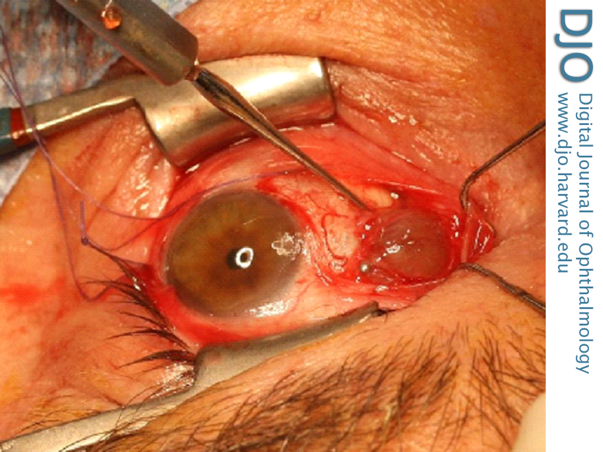
Figure 3
Intraoperative appearance of the chocolate-colored cyst filled with serosanguninous fluid, with strands of flaccid extraocular muscle straddling its surface.
Intraoperative appearance of the chocolate-colored cyst filled with serosanguninous fluid, with strands of flaccid extraocular muscle straddling its surface.
The lining of the cyst in the present case was composed of epithelial cells, and this limited the differential to epidermal cyst, conjunctival cyst, respiratory epithelial cyst, and apocrine gland cyst. The lack of keratin production and of squamous differentiation excluded the possibility of epidermal cyst. The lack of cilia and goblet cells made respiratory epithelial cyst unlikely reducing the differential to an apocrine gland cyst (sudoriferous cyst) or a conjunctival cyst. Apocrine gland cysts, which may (rarely) occur in the anterior portion of the orbit, are congenital in nature and thought to develop from entrapped epithelial cells destined to form the glands of Moll. Most conjunctival cysts develop secondary to implantation following ocular trauma or surgery. A primary, congenital form has been described that arises along the common sheath of the superior rectus muscle and levator palpebrae superioris muscle that is thought to result from misdirected cleavage of mesoderm.(2)
Development of a clinically significant giant conjunctival cyst as a complication of strabismus surgery has been reported, albeit rarely.(3-6) This complication has a reported incidence of 0.25% after strabismus procedures.(7) The purple/blue grayish hue of such cysts, noted in our patient, has been previously cited along with the frequent delay in growth, secondary strabismus, and clinical presentation for evaluation.(6,8)
Although thermal cauterization or intralesional injection with isopropyl alcohol have been successfully used to shrink some conjunctival cysts,(9,10) these techniques do not address the large secondary incommitant strabismus associated with many giant conjunctival cysts. Management by marsupialization of the cyst has been reported to correct a simple epithelial cyst adjacent to a muscle.(11) In cases such as the present one, where the cyst has encapsulated muscle fibers and is associated with strabismus, careful surgical excision remains the mainstay of therapy because it allows salvaging the healthy rectus muscle and recreating a new attachment to the globe.(6,8,9)
When examining a patient with an orbital mass with or without associated strabismus, it is vital to take into account the history of previous ocular surgery or trauma and to consider the possibility of a giant conjunctival cyst. The use of imaging modalities, such as orbital MRI or ultrasound, can help provide useful information regarding the consistency of the mass and its relationship to adjacent structures. An isolated cyst without adjacent bony erosion or soft tissue invasion is likely to be benign histologically, although it is not benign if it causes secondary strabismus, displacement, or compression of surrounding structures. Imaging in the present case provided invaluable insight into the cystic structure of the mass, and the extreme displacement of the medial rectus insertion; understanding this anatomical relationship prevented inadvertent loss of the medial rectus at the time of repair. Histopathological examination confirmed the nature of the lesion in this case, reassuring patient and surgeon alike.
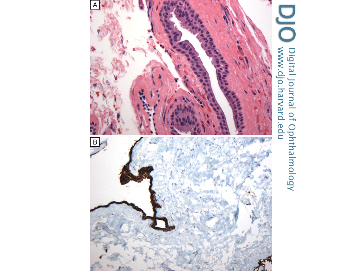
Figure 4
Histological characteristics of the mass. A, Hematoxylin and eosin stain showing a thin wall lined by a double layer of non-keratinizing cuboidal epithelium; there are no identifiable goblet cells (original magnification × 40). B, Immunohistochemistry for pan keratin highlighting the epithelial lining (original magnification × 20).
Histological characteristics of the mass. A, Hematoxylin and eosin stain showing a thin wall lined by a double layer of non-keratinizing cuboidal epithelium; there are no identifiable goblet cells (original magnification × 40). B, Immunohistochemistry for pan keratin highlighting the epithelial lining (original magnification × 20).
2. Rose GE, O’Donnell BA. Congenital orbital cysts associated with the common sheath of superior rectus and levator palpebrae superioris muscles. Ophthalmology 1995;102:135-8.
3. Metz HS, Searl S, Rosenberg P, Sterns G. Giant orbital cyst after strabismus surgery. J AAPOS 1999;3:185-7.
4. Cibis GW, Waeltermann JM. Muscle inclusion cyst as a complication of strabismus surgery. Am J Ophthalmol 1985;100:740-1.
5. Kushner BJ. Subconjunctival cysts as a complication of strabismus surgery. Arch Ophthalmol 1992;110:1243-5.
6. Song JJ, Finger PT, Kurli M, Wisnicki HJ, Iacob CE. Giant secondary conjunctival inclusion cysts: a late complication of strabismus surgery. Ophthalmology 2006;113:1049.e1041-2.
7. Guadilla AM, de Liaño PG, Merino P, Franco G. Conjunctival cysts as a complication after strabismus surgery. J Pediatr Ophthalmol Strabismus 2011;48:298-300.
8. Curtis TH, Stout AU, Drack AV, Durairaj VD. Giant orbital cysts after strabismus surgery. Am J Ophthalmol 2006;142:697-9.
9. Hawkins AS, Hamming NA. Thermal cautery as a treatment for conjunctival inclusion cyst after strabismus surgery. J AAPOS 2001;5:48-9.
10. Kothari M. A novel method for management of conjunctival inclusion cysts following strabismus surgery using isopropyl alcohol with paired injection technique. J AAPOS 2009;13:521-2.
11. Pereira LS, Hwang TN, McCulley TJ. Marsupialization of orbital conjunctival inclusion cysts related to strabismus surgery. J Pediatr Ophthalmol Strabismus 2009;46:180-1.